
Page 123 - Clinical Anatomy
P. 123
ECA2 7/18/06 6:42 PM Page 108
108 The abdomen and pelvis
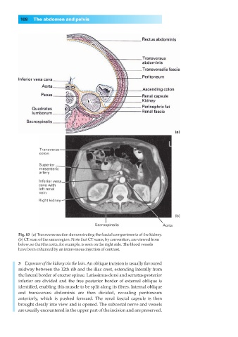
Fig. 83◊(a) Transverse section demonstrating the fascial compartments of the kidney.
(b) CT scan of the same region. Note that CT scans, by convention, are viewed from
below, so that the aorta, for example, is seen on the right side. The blood vessels
have been enhanced by an intravenous injection of contrast.
3◊◊Exposure of the kidney via the loin. An oblique incision is usually favoured
midway between the 12th rib and the iliac crest, extending laterally from
the lateral border of erector spinae. Latissimus dorsi and serratus posterior
inferior are divided and the free posterior border of external oblique is
identified, enabling this muscle to be split along its fibres. Internal oblique
and transversus abdominis are then divided, revealing peritoneum
anteriorly, which is pushed forward. The renal fascial capsule is then
brought clearly into view and is opened. The subcostal nerve and vessels
are usually encountered in the upper part of the incision and are preserved.