
Page 125 - Clinical Anatomy
P. 125
ECA2 7/18/06 6:42 PM Page 110
110 The abdomen and pelvis
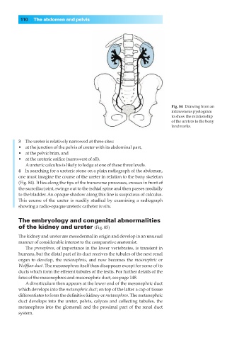
Fig. 84◊Drawing from an
intravenous pyelogram
to show the relationship
of the ureters to the bony
landmarks.
3◊◊The ureter is relatively narrowed at three sites:
•◊◊at the junction of the pelvis of ureter with its abdominal part,
•◊◊at the pelvic brim, and
•◊◊at the ureteric orifice (narrowest of all).
Aureteric calculus is likely to lodge at one of these three levels.
4◊◊In searching for a ureteric stone on a plain radiograph of the abdomen,
one must imagine the course of the ureter in relation to the bony skeleton
(Fig. 84). It lies along the tips of the transverse processes, crosses in front of
the sacroiliac joint, swings out to the ischial spine and then passes medially
to the bladder. An opaque shadow along this line is suspicious of calculus.
This course of the ureter is readily studied by examining a radiograph
showing a radio-opaque ureteric catheter in situ.
The embryology and congenital abnormalities
of the kidney and ureter (Fig. 85)
The kidney and ureter are mesodermal in origin and develop in an unusual
manner of considerable interest to the comparative anatomist.
The pronephros, of importance in the lower vertebrates, is transient in
humans, but the distal part of its duct receives the tubules of the next renal
organ to develop, the mesonephros, and now becomes the mesonephric or
Wolffian duct. The mesonephros itself then disappears except for some of its
ducts which form the efferent tubules of the testis. For further details of the
fates of the mesonephros and mesonephric duct, see page 148.
A diverticulum then appears at the lower end of the mesonephric duct
which develops into the metanephric duct; on top of the latter a cap of tissue
differentiates to form the definitive kidney or metanephros. The metanephric
duct develops into the ureter, pelvis, calyces and collecting tubules, the
metanephros into the glomeruli and the proximal part of the renal duct
system.