
Page 122 - Clinical Anatomy
P. 122
ECA2 7/18/06 6:42 PM Page 107
The urinary tract 107
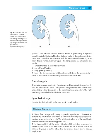
Fig. 82◊Variations in the
renal pelvis. (a) The
pelvis is buried within
the renal parenchyma—
pyelolithotomy difficult.
(b) The pelvis protrudes
generously—
pyelolithotomy easy.
(which is thus easily separated and left behind in performing a nephrec-
tomy). Medially, the fascia blends with the sheaths of the aorta and inferior
vena cava. Laterally it is continuous with the transversalis fascia. Only infe-
riorly does it remain relatively open — tracking around the ureter into the
pelvis.
The kidney has, in fact, three capsules:
1◊◊fascial (renal fascia);
2◊◊fatty (perinephric fat);
3◊◊true— the fibrous capsule which strips readily from the normal kidney
surface but adheres firmly to an organ that has been inflamed.
Blood supply
The renal artery derives directly from the aorta. The renal vein drains directly
into the inferior vena cava. The left renal vein passes in front of the aorta
immediately below the origin of the superior mesenteric artery. The right
renal artery passes behind the inferior vena cava.
Lymph drainage
Lymphatics drain directly to the para-aortic lymph nodes.
Clinical features
1◊◊Blood from a ruptured kidney or pus in a perinephric abscess first
distend the renal fascia, then force their way within the fascial compart-
ment downwards into the pelvis. The midline attachment of the renal fascia
prevents extravasation to the opposite side.
2◊◊In hypermobility of the kidney (‘floating kidney’), this organ can be
moved up and down in its fascial compartment but not from side to side. To
a lesser degree, it is in this plane that the normal kidney moves during
respiration.