
Page 148 - Clinical Anatomy
P. 148
ECA2 7/18/06 6:43 PM Page 133
The muscles of the pelvic floor and perineum 133
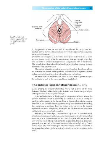
Fig. 97◊Levator ani—
inferior aspect. It forms
the ‘diaphragm of the
pelvis’.
3◊◊the posterior fibres are attached to the sides of the coccyx and to a
median fibrous raphe, which stretches between the apex of the coccyx and
the anorectal junction.
(Note that the coccygeus is in the same tissue plane as levator ani. It corre-
sponds almost exactly with the sacrospinous ligament, which it overlies,
and the latter is commonly regarded as a degenerate part of the muscle.
The muscle is well developed and the ligament is often missing in those
mammals with a mobile tail.)
The muscle acts as the principal support of the pelvic floor, has a sphinc-
ter action on the rectum and vagina and assists in increasing intra-abdomi-
nal pressure during defaecation, micturition and parturition.
Its deep aspect is related to the pelvic viscera and its perineal aspect
forms the inner wall of the ischiorectal fossa (see below).
The anterior (urogenital) perineum (Figs 98, 99)
A line joining the ischial tuberosities passes just in front of the anus.
Between this line and the ischiopubic inferior rami lies the urogenital part
of the perineum or the urogenital triangle.
Attached to the sides of this triangle is a tough fascial sheet termed the
perineal membrane which is pierced by the urethra in the male and by the
urethra and the vagina in the female. Deep to this membrane is the external
sphincter of the urethra consisting of voluntary muscle fibres surrounding
the membranous urethra; these are competent even when the internal
sphincter has been completely destroyed. In the female the superficial
sphincter is also pierced by the vagina.
Enclosing the deep aspect of the external sphincter is a second fascial
sheath (comprising areolar tissue on the deep aspect of levator ani), so that
this muscle is, in fact, contained within a fascial capsule which is termed the
deep perineal pouch. This pouch contains, in addition, the deep transverse
perineal muscles and, in the male, the two bulbo-urethral glands of Cowper
whose ducts pass forward to open into the bulbous urethra. Superficial to
the perineal membrane is the superficial perineal pouch which contains, in the
male: