
Page 154 - Clinical Anatomy
P. 154
ECA2 7/18/06 6:43 PM Page 139
The female genital organs 139
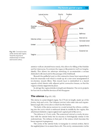
Fig. 102◊Coronal section
of the uterus and vagina.
Note the important
relationships of ureter
and uterine artery.
anterior wall are situated transversely; this allows for filling of the bladder
and for intercourse. In contrast, the rugae on the posterior wall run longitu-
dinally. This allows for sideways stretching to accommodate a rectum
distended with stool and for the passage of the fetal head.
Beneath the epithelial coat is a thin connective tissue layer separating it
from the muscular wall which is made up of a criss-cross arrangement of
involuntary muscle fibres. This muscle layer is ensheathed in a fascial
capsule which blends with adjacent pelvic connective tissues, so that the
vagina is firmly supported in place.
In old age the vagina shrinks in length and diameter. The cervix projects
far less into it so that the fornices all but disappear.
The uterus (Figs 101, 102)
The uterus is a pear-shaped organ, 3in (7.5cm) in length, made up of the
fundus, body and cervix. The Fallopian (uterine) tubes enter into each supero-
lateral angle (the cornu) above which lies the fundus.
The body of the uterus narrows to a waist termed the isthmus, continu-
ing into the cervix which is embraced about its middle by the vagina; this
attachment delimits a supravaginal and vaginal part of the cervix.
The isthmus is 1.5mm wide. The anatomical internal os marks its junc-
tion with the uterine body but its mucosa is histologically similar to the
endometrium. The isthmus is that part of the uterus which becomes the
lower segment in pregnancy.
The cavity of the uterine body is triangular in coronal section, but in
the sagittal plane it is no more than a slit. This cavity communicates via the