
Page 151 - Clinical Anatomy
P. 151
ECA2 7/18/06 6:43 PM Page 136
136 The abdomen and pelvis
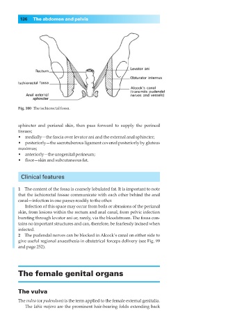
Fig. 100◊The ischiorectal fossa.
sphincter and perianal skin, then pass forward to supply the perineal
tissues;
•◊◊medially—the fascia over levator ani and the external anal sphincter;
•◊◊posteriorly—the sacrotuberous ligament covered posteriorly by gluteus
maximus;
•◊◊anteriorly—the urogenital perineum;
•◊◊floor—skin and subcutaneous fat.
Clinical features
1◊◊The content of the fossa is coarsely lobulated fat. It is important to note
that the ischiorectal fossae communicate with each other behind the anal
canal—infection in one passes readily to the other.
Infection of this space may occur from boils or abrasions of the perianal
skin, from lesions within the rectum and anal canal, from pelvic infection
bursting through levator ani or, rarely, via the bloodstream. The fossa con-
tains no important structures and can, therefore, be fearlessly incised when
infected.
2◊◊The pudendal nerves can be blocked in Alcock’s canal on either side to
give useful regional anaesthesia in obstetrical forceps delivery (see Fig. 99
and page 252).
The female genital organs
The vulva
The vulva (or pudendum) is the term applied to the female external genitalia.
The labia majora are the prominent hair-bearing folds extending back