
Page 153 - Clinical Anatomy
P. 153
ECA2 7/18/06 6:43 PM Page 138
138 The abdomen and pelvis
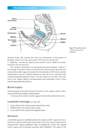
Fig. 101◊Sagittal section
of the uterus and its
relations.
perineal body), the rectum and then the peritoneum of the pouch of
Douglas which covers the upper quarter of the posterior vaginal wall.
•◊◊Laterally — levator ani, pelvic fascia and the ureters, which lie immedi-
ately above the lateral fornices.
The amateur abortionist (or inexperienced gynaecologist) without a
knowledge of anatomy fails to realize that the uterus passes upwards and
forwards from the vagina; he pushes the instrument or IUCD (intra-uterine
contraceptive device), which he intends to enter the cervix, directly back-
wards through the posterior fornix. To make matters worse, this is the only
part of the vagina which is intraperitoneal; the peritoneal cavity is thus
entered and peritonitis follows.
Blood supply
Arterial supply is from the internal iliac artery via its vaginal, uterine, inter-
nal pudendal and middle rectal branches.
Avenous plexus drains via the vaginal vein into the internal iliac vein.
Lymphatic drainage (see Fig. 105)
•◊◊Upper third to the external and internal iliac nodes.
•◊◊Middle third to the internal iliac nodes.
•◊◊Lower third to the superficial inguinal nodes.
Structure
A stratified squamous epithelium lines the vagina and the vaginal cervix; it
contains no glands and is lubricated partly by cervical mucus and partly by
desquamated vaginal epithelial cells. In nulliparous women the vaginal
wall is rugose, but it becomes smoother after childbirth. The rugae of the