
Page 149 - Clinical Anatomy
P. 149
ECA2 7/18/06 6:43 PM Page 134
134 The abdomen and pelvis
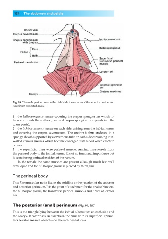
Fig. 98◊The male perineum—on the right side the muscles of the anterior perineum
have been dissected away.
1◊◊the bulbospongiosus muscle covering the corpus spongiosum which, in
turn, surrounds the urethra (the distal corpus spongiosum expands into the
glans penis);
2◊◊the ischiocavernosus muscle on each side, arising from the ischial ramus
and covering the corpus cavernosum. The urethra is thus enclosed in a
spongy sheath supported by a cavernous tube on each side containing thin-
walled venous sinuses which become engorged with blood when erection
occurs;
3◊◊the superficial transverse perineal muscle, running transversely from
the perineal body to the ischial ramus. It is of no functional importance but
is seen during perineal excision of the rectum.
In the female the same muscles are present although much less well
developed and the bulbospongiosus is pierced by the vagina.
The perineal body
This fibromuscular node lies in the midline at the junction of the anterior
and posterior perineum. It is the point of attachment for the anal sphincters,
the bulbospongiosus, the transverse perineal muscles and fibres of levator
ani.
The posterior (anal) perineum (Figs 99, 100)
This is the triangle lying between the ischial tuberosities on each side and
the coccyx. It comprises, in essentials, the anus with its superficial sphinc-
ters, levator ani and, at each side, the ischiorectal fossa.