
Page 150 - Clinical Anatomy
P. 150
ECA2 7/18/06 6:43 PM Page 135
The muscles of the pelvic floor and perineum 135
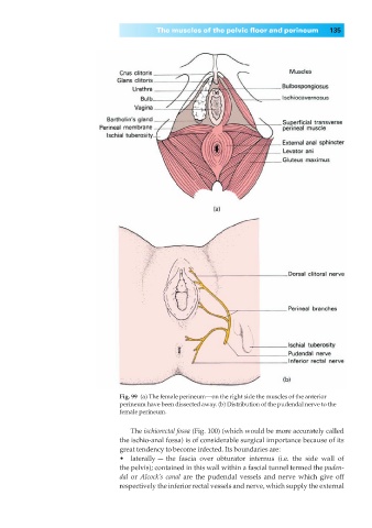
Fig. 99◊(a) The female perineum—on the right side the muscles of the anterior
perineum have been dissected away. (b) Distribution of the pudendal nerve to the
female perineum.
The ischiorectal fossa (Fig. 100) (which would be more accurately called
the ischio-anal fossa) is of considerable surgical importance because of its
great tendency to become infected. Its boundaries are:
•◊◊laterally — the fascia over obturator internus (i.e. the side wall of
the pelvis); contained in this wall within a fascial tunnel termed the puden-
dal or Alcock’s canal are the pudendal vessels and nerve which give off
respectively the inferior rectal vessels and nerve, which supply the external