
Page 181 - Clinical Anatomy
P. 181
ECA3 7/18/06 6:45 PM Page 166
166 The upper limb
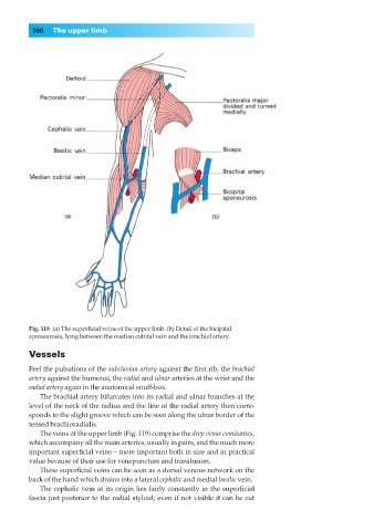
Fig. 119◊(a) The superficial veins of the upper limb. (b) Detail of the bicipital
aponeurosis, lying between the median cubital vein and the brachial artery.
Vessels
Feel the pulsations of the subclavian artery against the first rib, the brachial
artery against the humerus, the radial and ulnar arteries at the wrist and the
radial artery again in the anatomical snuff-box.
The brachial artery bifurcates into its radial and ulnar branches at the
level of the neck of the radius and the line of the radial artery then corre-
sponds to the slight groove which can be seen along the ulnar border of the
tensed brachioradialis.
The veins of the upper limb (Fig. 119) comprise the deep venae comitantes,
which accompany all the main arteries, usually in pairs, and the much more
important superficial veins— more important both in size and in practical
value because of their use for venepuncture and transfusion.
These superficial veins can be seen as a dorsal venous network on the
back of the hand which drains into a lateral cephalic and medial basilic vein.
The cephalic vein at its origin lies fairly constantly in the superficial
fascia just posterior to the radial styloid; even if not visible it can be cut