
Page 288 - Clinical Anatomy
P. 288
ECA5 7/18/06 6:50 PM Page 273
The tongue and floor of the mouth 273
embryological origin of the thyroid (see page 286). Immediately in front of
the sulcus lie a row of large vallate papilliae.
The under aspect of the tongue bears the median frenulum linguae;
the mucosa is thin on this surface and the lingual veins can thus be seen
on either side of the frenulum. The lingual nerve and the lingual artery
are medial to the vein but not visible. More laterally can be seen a fringed
fold of mucous membrane termed the plica fimbriata. On either side of
the base of the frenulum can be seen the orifice of the submandibular
duct on its papilla. Inspect this in a mirror and note the discharge of saliva
when you press on your submandibular gland just below the angle of
the jaw.
Structure
The thick stratified squamous mucosa of the dorsum of the tongue bears
papillae over the anterior two-thirds back as far as the sulcus terminalis.
These papillae (particularly the vallate) bear the taste buds. The posterior
one-third has no papillae but carries numerous lymphoid nodules which,
with the palatine tonsils and adenoids, make up the lymphoid ring of
Waldeyer.
Small glands are scattered throughout the submucosa of the dorsum;
these are predominantly serous anteriorly and mucous posteriorly.
The tongue is divided by a median vertical fibrous septum, as indicated
on the dorsum by a shallow groove. On each side of this septum are the
intrinsic and extrinsic muscles of the tongue (Fig. 197).
The intrinsic muscles are disposed in vertical, longitudinal and trans-
verse bundles; they alter the shape of the tongue.
The extrinsic muscles move the tongue as a whole. They pass to the
tongue from the symphysis of the mandible, the hyoid, styloid process and
the soft palate, respectively the genioglossus, hyoglossus, styloglossus and
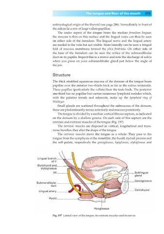
Fig. 197◊Lateral view of the tongue, its extrinsic muscles and its nerves.