
Page 289 - Clinical Anatomy
P. 289
ECA5 7/18/06 6:50 PM Page 274
274 The head and neck
palatoglossus. The functions of the individual extrinsic muscles can be
deduced from their relative positions (Fig. 197). Genioglossus protrudes
the tongue, styloglossus retracts it and hyoglossus depresses it. Palatoglos-
sus is, in fact, a palatal muscle and helps to narrow the oropharynx in
swallowing.
Blood supply
Blood is supplied from the lingual branch of the external carotid artery.
There is little cross-circulation across the median raphe, which is therefore a
relatively avascular plane.
Lymph drainage (Fig. 198)
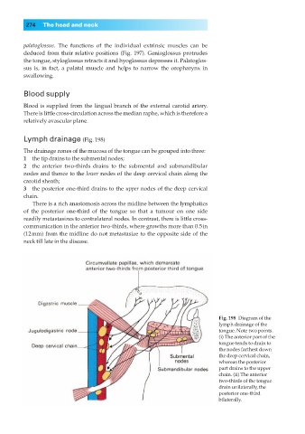
The drainage zones of the mucosa of the tongue can be grouped into three:
1◊◊the tip drains to the submental nodes;
2◊◊the anterior two-thirds drains to the submental and submandibular
nodes and thence to the lower nodes of the deep cervical chain along the
carotid sheath;
3◊◊the posterior one-third drains to the upper nodes of the deep cervical
chain.
There is a rich anastomosis across the midline between the lymphatics
of the posterior one-third of the tongue so that a tumour on one side
readily metastasizes to contralateral nodes. In contrast, there is little cross-
communication in the anterior two-thirds, where growths more than 0.5in
(12mm) from the midline do not metastasize to the opposite side of the
neck till late in the disease.
Fig. 198◊Diagram of the
lymph drainage of the
tongue. Note two points.
(i) The anterior part of the
tongue tends to drain to
the nodes farthest down
the deep cervical chain,
whereas the posterior
part drains to the upper
chain. (ii) The anterior
two-thirds of the tongue
drain unilaterally, the
posterior one-third
bilaterally.