
Page 356 - Clinical Anatomy
P. 356
ECA6 7/18/06 6:54 PM Page 341
The brain 341
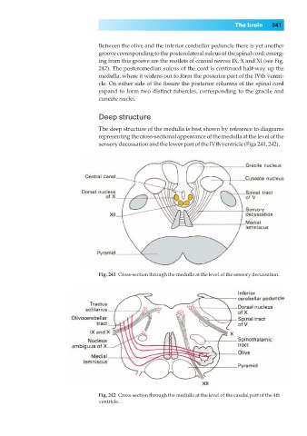
Between the olive and the inferior cerebellar peduncle there is yet another
groove corresponding to the posterolateral sulcus of the spinal cord; emerg-
ing from this groove are the rootlets of cranial nerves IX, X and XI (see Fig.
242). The posteromedian sulcus of the cord is continued half-way up the
medulla, where it widens out to form the posterior part of the IVth ventri-
cle. On either side of the fissure the posterior columns of the spinal cord
expand to form two distinct tubercles, corresponding to the gracile and
cuneate nuclei.
Deep structure
The deep structure of the medulla is best shown by reference to diagrams
representing the cross-sectional appearance of the medulla at the level of the
sensory decussation and the lower part of the IVth ventricle (Figs 241, 242).
Fig. 241◊Cross-section through the medulla at the level of the sensory decussation.
Fig. 242◊Cross-section through the medulla at the level of the caudal part of the 4th
ventricle.