
Page 360 - Clinical Anatomy
P. 360
ECA6 7/18/06 6:54 PM Page 345
The brain 345
are the two cerebral peduncles, which emerge from the substance of the cere-
bral hemisphere and pass downwards and medially, connecting the inter-
nal capsule to the pons. The fibres of the 3rd nerves emerge between the
two cerebral peduncles in the interpeduncular fossa. Viewed from the lateral
aspect, the midbrain can be seen to consist of three distinct portions: the
basis pedunculi ventrally, the midbrain tegmentum centrally and the tectum
dorsally. The trochlear nerve (IV), the optic tract and the posterior cerebral
artery wind around this aspect of the midbrain. The dorsal surface of the
midbrain presents the four colliculi (or corpora quadrigemini) and the supe-
rior medullary velum between the two superior cerebellar peduncles. The
pineal gland rests between the two superior colliculi and is attached by a
stalk to the posterior dorsal thalamus. It secretes melatonin and has an
important role in setting the circadian rhythm.
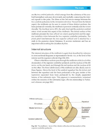
Internal structure
The internal structure of the midbrain is again best described by reference
to cross-sectional diagrams at representative levels: viz. at the level of the
inferior and the superior colliculi (Figs 244, 245).
Observe that these sections pass through the midbrain at the level of the
decussation of the superior cerebellar peduncle and the nucleus of the 4th
nerve, on the one hand, and through the red nucleus and the nucleus of III
on the other. The three subdivisions of the midbrain are also clearly seen in
these figures. Above the level of the cerebral aqueduct lies the tectum and
between the aqueduct and the basis pedunculi is the grey matter of the
tegmentum separated from basis pedunculi by the deeply pigmented
lamina of the substantia nigra. This pigment is neuromelanin, contained
within the neurons of the substantia nigra. (For its relationship to Parkin-
son’s disease, see page 360.)
Fig. 244◊The midbrain—level of the inferior colliculus and decussation of the
superior cerebellar peduncle.