
Page 359 - Clinical Anatomy
P. 359
ECA6 7/18/06 6:54 PM Page 344
344 The central nervous system
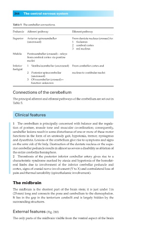
Table 5◊The cerebellar connections.
Peduncle Afferent pathway Efferent pathway
Superior Anterior spinocerebellar From dentate nucleus (crossed) to:
(uncrossed) 1◊◊thalamus
2◊◊cerebral cortex
3◊◊red nucleus
Middle Pontocerebellar (crossed)—relays
from cerebral cortex via pontine
nuclei
Inferior 1◊◊Vestibulocerebellar (uncrossed) From cerebellar cortex and
fastigial
2◊◊Posterior spinocerebellar nucleus to vestibular nuclei
◊◊1(uncrossed)
3◊◊Olivocerebellar (crossed)—
◊◊1function unknown
Connections of the cerebellum
The principal afferent and efferent pathways of the cerebellum are set out in
Table 5.
Clinical features
1◊◊The cerebellum is principally concerned with balance and the regula-
tion of posture, muscle tone and muscular co-ordination; consequently,
cerebellar lesions result in some disturbance of one or more of these motor
functions in the form of an unsteady gait, hypotonia, tremor, nystagmus
and dysarthria. Lesions of the cerebellum give rise to symptoms and signs
on the same side of the body. Destruction of the dentate nucleus or the supe-
rior cerebellar peduncle results in almost as severe a disability as ablation of
the entire cerebellar hemisphere.
2◊◊Thrombosis of the posterior inferior cerebellar artery gives rise to a
characteristic syndrome marked by ataxia and hypotonia of the homolat-
eral limbs due to involvement of the inferior cerebellar peduncle and
cortex, signs of cranial nerve involvement (V to X) and contralateral loss of
pain and thermal sensibility (spinothalamic involvement).
The midbrain
The midbrain is the shortest part of the brain stem; it is just under 1in
(25mm) long and connects the pons and cerebellum to the diencephalon.
It lies in the gap in the tentorium cerebelli and is largely hidden by the
surrounding structures.
External features (Fig. 240)
The only parts of the midbrain visible from the ventral aspect of the brain