
Page 361 - Clinical Anatomy
P. 361
ECA6 7/18/06 6:54 PM Page 346
346 The central nervous system
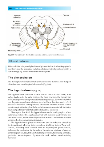
Fig. 245◊The midbrain—level of the superior colliculus and the red nucleus.
Clinical features
When calcified, the pineal gland is easily identified on skull radiographs. It
may then give the important radiological sign of lateral displacement by a
space-occupying lesion of the cerebral hemisphere.
The diencephalon
The diencephalon comprises the hypothalamus and thalamus. It is that part
of the brain surrounding the 3rd ventricle (Fig. 246).
The hypothalamus (Fig. 246)
The hypothalamus forms the floor of the 3rd ventricle. It includes, from
before backwards, the optic chiasma, the tuber cinereum, the infundibular
stalk(leading down to the posterior lobe of the pituitary), the mamillary bodies
and the posterior perforated substance. In each of these there is a number of cell
masses or nuclei and a fibre pathway—the medial forebrain bundle—which
runs throughout the length of the hypothalamus and serves to link it with the
midbrain posteriorly and the basal forebrain areas anteriorly.
Sherrington described the hypothalamus as the head ganglion of the
autonomic system. It is largely concerned with autonomic activity and can
be divided into a posteromedial sympathetic area and an anterolateral area
concerned with parasympathetic activity.
The hypothalamus plays an important part in endocrine control by
the formation of releasing factors or release-inhibiting factors. These sub-
stances, following their secretion into the hypophyseal portal vessels,
influence the production by the cells of the anterior pituitary of adreno-
corticotrophin (ACTH), follicle-stimulating hormone, luteinizing hormone,
prolactin, somatotrophin, thyrotrophin and melanocyte-stimulating
hormone.