
Page 358 - Clinical Anatomy
P. 358
ECA6 7/18/06 6:54 PM Page 343
The brain 343
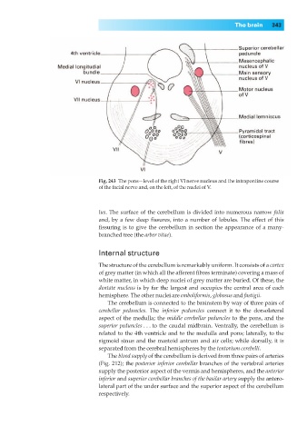
Fig. 243◊The pons—level of the right VI nerve nucleus and the intrapontine course
of the facial nerve and, on the left, of the nuclei of V.
lus. The surface of the cerebellum is divided into numerous narrow folia
and, by a few deep fissures, into a number of lobules. The effect of this
fissuring is to give the cerebellum in section the appearance of a many-
branched tree (the arbor vitae).
Internal structure
The structure of the cerebellum is remarkably uniform. It consists of a cortex
of grey matter (in which all the afferent fibres terminate) covering a mass of
white matter, in which deep nuclei of grey matter are buried. Of these, the
dentate nucleus is by far the largest and occupies the central area of each
hemisphere. The other nuclei are emboliformis, globosus and fastigii.
The cerebellum is connected to the brainstem by way of three pairs of
cerebellar peduncles. The inferior peduncles connect it to the dorsolateral
aspect of the medulla; the middle cerebellar peduncles to the pons, and the
superior peduncles ... to the caudal midbrain. Ventrally, the cerebellum is
related to the 4th ventricle and to the medulla and pons; laterally, to the
sigmoid sinus and the mastoid antrum and air cells; while dorsally, it is
separated from the cerebral hemispheres by the tentorium cerebelli.
The blood supply of the cerebellum is derived from three pairs of arteries
(Fig. 212); the posterior inferior cerebellar branches of the vertebral arteries
supply the posterior aspect of the vermis and hemispheres, and the anterior
inferior and superior cerebellar branches of the basilar artery supply the antero-
lateral part of the under surface and the superior aspect of the cerebellum
respectively.