
Page 362 - Clinical Anatomy
P. 362
ECA6 7/18/06 6:54 PM Page 347
The brain 347
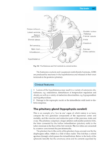
Fig. 246◊The thalamus and 3rd ventricle in coronal section.
The hormones oxytocin and vasopressin (anti-diuretic hormone, ADH)
are produced by neurones in the hypothalamus and released at their axon
terminals in the posterior pituitary.
Clinical features
1◊◊Lesions of the hypothalamus may result in a variety of autonomic dis-
turbances, e.g. somnolence, disturbances of temperature regulation and
obesity, as well as a variety of endocrine abnormalities, e.g. hypogonadism
and hypothyroidism.
2◊◊Damage to the supraoptic nuclei or the infundibular stalk leads to dia-
betes insipidus.
The pituitary gland (hypophysis cerebri)
This is an example of a ‘two in one’ organ of which nature is so keen;
compare the two glandular components of the suprarenal cortex and
medulla, and the exocrine and endocrine parts of the pancreas, testis and
ovary. The pituitary comprises a larger anterior and smaller posterior lobe,
the latter connected by the hollow infundibulum (pituitary stalk) to the
tuber cinereum in the floor of the 3rd ventricle. The two lobes are connected
by a narrow zone termed the pars intermedia.
The pituitary lies in the cavity of the pituitary fossa covered over by the
diaphragma sellae, which is a fold of dura mater. This fold has a central
aperture through which passes the infundibulum. Below is the body of the
sphenoid, laterally lies the cavernous sinus and its contents separated by