
Page 405 - Clinical Anatomy
P. 405
ECA6 7/18/06 6:54 PM Page 390
390 The central nervous system
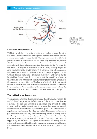
Fig. 272◊The right
fundus oculi as seen
through an
ophthalmoscope.
Contents of the eyeball
Within the eyeball are found: the lens, the aqueous humour and the vitre-
ous body. The lens is biconvex and is placed between the vitreous and the
aqueous humour, just behind the iris. The aqueous humour is a filtrate of
plasma secreted by the vessels of the iris and ciliary body into the posterior
chamber of the eye (i.e. the space between the lens and the iris). From here it
passes through the pupillary aperture into the anterior chamber (between the
cornea and the iris) and is re-absorbed into the ciliary veins by way of the
sinus venosus sclerae (or canal of Schlemm). The vitreous body, which occupies
the posterior four-fifths of the eyeball, is a thin transparent gel contained
within a delicate membrane — the hyaloid membrane — and pierced by the
lymph-filled hyaloid canal. The anterior part of the hyaloid membrane is
thickened, receives attachments from the ciliary processes and gives rise to
the suspensory ligament of the lens. This ligament is attached to the capsule of
the lens in front of its equator and serves to retain it in position. It is relaxed
by contraction of the radial fibres of the ciliary muscle and so allows the
lens to assume a more convex form in accommodation (close reading).
The orbital muscles (Fig. 262)
These are the levator palpebrae superioris and the extra-ocular muscles; the
medial, lateral, superior and inferior recti and the superior and inferior
obliques. The four recti arise from a tendinous ring around the optic
foramen and the medial part of the superior orbital fissure and are inserted
into the sclera anterior to the equator of the eyeball. The lateral rectus is
supplied by the 6th nerve, the others by the 3rd. The superior oblique arises
just above the tendinous ring and is inserted by means of a long tendon
which loops around a fibrous pulley on the medial part of the roof of the
orbit into the sclera just lateral to the insertion of the superior rectus. It is
supplied by the 4th nerve. The inferior oblique passes like a sling from its
origin on the medial side of the orbit around the undersurface of the eye
to insert into the sclera between the superior and lateral recti; it is supplied
by III.