
Page 403 - Clinical Anatomy
P. 403
ECA6 7/18/06 6:54 PM Page 388
388 The central nervous system
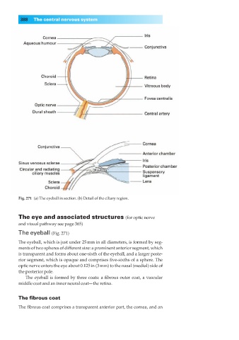
Fig. 271◊(a) The eyeball in section. (b) Detail of the ciliary region.
The eye and associated structures (for optic nerve
and visual pathway see page 365)
The eyeball (Fig. 271)
The eyeball, which is just under 25mm in all diameters, is formed by seg-
ments of two spheres of different size: a prominent anterior segment, which
is transparent and forms about one-sixth of the eyeball, and a larger poste-
rior segment, which is opaque and comprises five-sixths of a sphere. The
optic nerve enters the eye about 0.125in (3mm) to the nasal (medial) side of
the posterior pole.
The eyeball is formed by three coats: a fibrous outer coat, a vascular
middle coat and an inner neural coat—the retina.
The fibrous coat
The fibrous coat comprises a transparent anterior part, the cornea, and an