
Page 16 - The Netter Collection of Medical Illustrations - Integumentary System_ Volume 4 ( PDFDrive )
P. 16
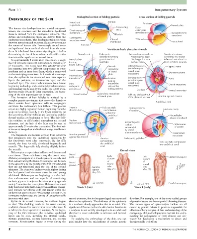
Plate 1-1                                                                                             Integumentary System

                                                      Midsagittal section of folding gastrula    Cross section of folding gastrula
       EMBRYOLOGY OF THE SKIN
                                                 Notochord                  Amnion                  Amnion
                                                 in gastrula                                                            Neural plate
       The human skin develops from two special embryonic                         Connecting   Extra-
       tissues,  the  ectoderm  and  the  mesoderm.  Epidermal   Oropharyngeal    stalk        embryonic
       tissue  is  derived  from  the  embryonic  ectoderm.  The   membrane     Allantois      mesoderm                 Intraembryonic
       dermis  and  subcutaneous  tissue  are  derived  from  the   Cardiogenic                                         mesoderm
       embryonic mesoderm. The developmental interactions   mesoderm          Cloacal          Yolk sac
       between mesoderm and ectoderm ultimately determine                     membrane                                  Notochord
       the  nature  of  human  skin.  Interestingly,  neural  tissue   Yolk sac
       and epidermal tissue are both derived from the ecto-                 Vertebrate body plan after 4 weeks
       derm. It is believed that calcium signaling is critical in
       determining the fate of the ectoderm and its differentia-  Neural crest  Embryonic  Intermediate mesoderm:  Somite sclerotome
       tion into either epidermis or neural tissue.                   endoderm forming        Nephrogenic ridge    surrounds the neural
         At approximately 4 weeks after conception, a single   Neural plate  gastrointestinal     Nephrogenic cord  tube and notochord to
       layer of ectoderm is present, surrounding a thicker layer   forming  (gut) tube        Genital ridge        form vertebral column
       of  mesoderm.  Two  weeks  later,  this  ectodermal  layer   neural tube  Somatic  Splanchnopleure             Spinal nerve
       has separated into two different components: an outer                mesoderm      (endoderm plus
       periderm and an inner basal layer, which is connected   Somite       of lateral    lateral plate               Dermomyotome
       to the underlying mesoderm. At 8 weeks after concep-  Intermediate   plate         mesoderm)
       tion, the epidermis has developed into three separate   mesoderm     Amnion        Somatopleure                Aorta
       layers:  the  periderm,  an  intermediate  layer,  and  the          tucking       (ectoderm plus
       basal cell layer. The dermal subcutaneous tissue is now   Intra-     around the    lateral plate
       beginning to develop, and a distinct dermal subcutane-  embryonic    sides of the  mesoderm)                   Dorsal mesentery
       ous boundary can be seen by the end of the eighth week.   coelom     folding embryo      Gut tube
       Between weeks 10 and 15 after conception, the begin-                                                           Ventral mesentery
       ning of the skin appendages can be seen.     Notochord        Splanchnic mesoderm  Yolk sac (stalk just out
         The  formation  of  hair  follicles  is  initiated  by  a   of lateral plate     of the plane of section)  Amnion against chorion
       complex genetic mechanism that causes the dermis to
       direct  certain  basal  epidermal  cells  to  congregate                                              Umbilical cord
       and  form  the  rudimentary  hair  follicle.  This  process
       occurs in a highly organized fashion beginning from the   Hepatic  Yolk sac stalk  Dermomyotome            Amniotic cavity
                                                 diverticulum
                                                                          and allantois
       scalp and working caudally to the lower extremity. At                 within the   of somite
       the same time, the hair follicles are developing and the                  umbilical                             Neural tube
       dermal papillae are beginning to form. The hair folli-                     cord                                 above notochord
       cles  continue  to  differentiate  throughout  the  second   Septum
       trimester,  and  the  hair  of  the  fetus  can  be  seen  at   transversum      Intraembryonic                    Sclerotome
                                                                                        coelom
       approximately 20 weeks after conception. This first hair                         surrounded                        of somite
       is known as lanugo hair and is almost always shed before                         by lateral
       delivery.                                                                        plate mesoderm                   Intermediate
         The fingernails and toenails develop from ectoderm   Amnion pressed                                             mesoderm
       that  invaginates  into  the  underlying  mesoderm  by     against the chorion  Amnion  Embryonic gut tube
       the  fourteenth  week  after  conception.  By  the  fifth       surrounding                              Yolk sac stalk compressed
       month,  the  fetus  has  fully  developed  fingernails  and     the umbilical                            into umbilical cord
       toenails.  The  fingernails  fully  develop  slightly  before   cord
       the toenails.                                                                 Dorsal views
         Melanocytes are specialized cells derived from neural
       crest  tissue.  These  cells  form  along  the  neural  tube.   Neural  Early                Late
       Melanocytes migrate in a specific pattern laterally and   plate      closure                 closure           Cranial neuropore
       then outward along the trunk. Melanocytes can be seen                of                      of
       in the epidermis by the middle of the first trimester, but   Neural  neural                  neural
                                                 groove
       they  are  not  functional  until  the  end  of  the  second   Somites  tube                 tube
       trimester. The density of melanocytes is highest during   appear     (day 21)                (day 22)
       the  fetal  period  and  decreases  thereafter  until  young   (day 20)
       adulthood.  Melanocytes  are  beginning  to  make  their
       first  melanosomes  and  are  capable  of  transferring
       melanin pigment to adjacent keratinocytes by approxi-
       mately 5 months after conception. Melanocytes are not       1.8 mm                       2.0-2.1 mm            Caudal neuropore
       fully functional until birth. Langerhans cells are special-  Week 3 (late)             Week 4 (early)
       ized immune surveillance cells that appear within the
       epidermis at approximately 40 days after conception. In
       contrast to melanocytes, the density of Langerhans cells
       increases with time.                      second trimester, first in the appendageal structures and   disorders. For example, one of the more studied groups
         By late in the second trimester, the periderm begins   then in the epidermis. The thickness of the epidermis   of genetic diseases are the congenital blistering diseases.
       to  shed.  This  shedding  results  in  the  vernix  caseosa,     in a newborn closely approaches that in an adult. The   The  various  types  of  epidermolysis  bullosa  are  all
       a whitish, cheese-like material that covers the fetus. It   significant difference is that the skin barrier function in   caused  by  genetic  defects  in  proteins  responsible  for
       is believed to have a protective function. At the begin-  a newborn is not as fully developed as in an adult and   adhesion of keratinocytes. A firm understanding of the
       ning  of  the  third  trimester,  the  individual  epidermal   therefore is more vulnerable to infection and external   embryology of skin development is essential for under-
       layers  can  be  seen,  including  the  stratum  basale,     insults.               standing  the  pathogenesis  of  these  diseases  and  ulti-
       stratum  granulosum,  stratum  spinosum,  and  stratum   By  studying  the  embryology  of  the  skin,  one  can     mately  for  developing  a  mechanism  to  detect  and
       corneum.  Keratinization  begins  to  occur  during  the   gain  insight  into  the  mechanisms  of  certain  genetic   therapeutically treat them.

       2                                                                                     THE NETTER COLLECTION OF MEDICAL ILLUSTRATIONS