
Page 18 - The Netter Collection of Medical Illustrations - Integumentary System_ Volume 4 ( PDFDrive )
P. 18
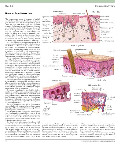
Plate 1-3                                                                                             Integumentary System

                                                             Glabrous skin                                Hairy skin
       NORMAL SKIN HISTOLOGY                                      Dermal papilla   Sweat gland Hair
                                                 Epidermis                                                         Hair follicle
       The  integumentary  system  is  composed  of  multiple   Krause                                             Merkel disc
       subunits that work in unison. The skin and its appenda-  end bulb
       geal  structures  make  up  the  integumentary  system.                                                     Free nerve ending
       There  are  three  main  layers  to  the  skin:  epidermis,   Free nerve
       dermis, and subcutaneous tissue. Within the epidermis,   ending
       the principal skin cell is the keratinocyte. Other cells   Meissner                                         Sebaceous gland
       found  in  the  epidermis  include  melanocytes,  Merkel   corpuscle
       cells, and Langerhans cells. The main cell type found                                                       Nerve plexus
       within  the  dermis  is  the  fibroblast.  Fibroblasts  make   Merkel disc                                  around hair
       collagen, which forms the mechanical support for the   Free nerve                                           follicle
       skin. The dermis is a region of high vascularity. The   ending                                              Ruffini terminals
       subcutaneous  fat  tissue  is  found  directly  beneath
       the dermis and is composed primarily of adipocytes.  Pacinian                                               Pacinian corpuscle
         The  normal  human  epidermis  varies  extensively  in   corpuscle
       thickness in different regions of the body. It is thickest
       on  the  back  and  thinnest  on  the  eyelids  and  on  the               Strata of epidermis
       scrotal skin. The epidermis can be subdivided into five
       components: stratum basale, stratum spinosum, stratum   Hair shaft             Langerhans cells
       granulosum,  stratum  lucidum,  and  stratum  corneum.                                                             Sweat duct
       The stratum lucidum is found only on the skin of the
       palms and soles. Each layer of the epidermis has impor-  Corneum
       tant anatomical and physiological functions.          Lucidum
         The stratum basale is the deepest layer. It consists of
       cuboidal epithelium sitting atop a basement membrane   Granulosum
       zone.  The  stratum  basale  contains  the  proliferating   Spinosum
       keratinocytes, which are constantly undergoing replica-
       tion to replace the overlying epidermis. It takes approx-  Basale or Germinativum
       imately 28 days for a basal keratinocyte to progress to
       the outermost layer of the stratum corneum. Melano-     Dermis
       cytes  and  Merkel  cells  can  also  be  found  within  the
       stratum basale. Melanocytes are pigment-forming cells;
       they  transfer  their  pigment  to  neighboring  keratino-
       cytes. Merkel cells are modified nerve endings and have         Basement membrane
       been found to be important as mechanoreceptors.
         The stratum spinosum is many cell layers thick and                                       Melanocytes  Merkel cells
       is  recognized  by  the  intercellular  connections  among   Glabrous skin
       adjacent keratinocytes, which are seen on light micros-
       copy as tiny spines. From the lower to the upper layers                                       Hair-bearing skin
       of  the  stratum  spinosum,  the  keratinocytes  progres-
       sively become flatter in appearance.                                                              Papillary loops
         The stratum granulosum is recognized by the large                             Epidermis         of dermal papillae
       number of basophilic keratohyalin granules within its
       keratinocytes. This stratum is typically 2 to 4 cell layers                   Papillary
       thick. The keratohyalin granules are composed primar-                         dermis
       ily of the protein profilaggrin; they vary from 1 to 4 µm                                                          Superficial
       in diameter. Profilaggrin is the precursor to filaggrin,                                                           plexus
       an essential protein that is required for the integrity of                    Reticular
       the overlying epidermis.                    Epidermis                         dermis
         The stratum lucidum occurs only in the skin of the   Dermis
       palms and soles. It is composed of a translucent eosino-                                                           Deep
       philic layer. The stratum lucidum is made up of tightly                                                            dermal
       packed squamous keratinocytes.                                                                                     plexus
         The stratum corneum, the outermost layer of skin, is                   Branches from
       made up of anucleate, cornified keratinocytes. Kerati-                   subcutaneous plexus                 Musculocutaneous
       nization (cornification) is a complex process that results                       Arteriovenous shunts        artery and vein
       in  the  appearance  of  the  stratum  corneum.  As  cells
       progress up the stratum corneum, they are shed in the
       process known as desquamation.
         The dermis is primarily composed of collagen, which
       is  produced  by  fibroblasts.  This  portion  of  the  skin   into two regions, called the papillary and the reticular   The subcutaneous tissue is composed of adipocytes.
       contains a highly vascular network that is responsible   portions.  The  papillary  dermis  is  juxtaposed  to  the   This tissue’s main functions are storage of energy, insu-
       for the nutrition of the skin and for thermoregulation.   overlying epidermis and interdigitates with it. The pap-  lation,  and  cushioning.  The  adipocytes  are  closely
       This  network  includes  a  deep  dermal  plexus  and  a   illary dermis and the epidermis are connected by the   packed  in  a  connective  tissue  septum  with  associated
       superficial plexus. The superficial plexus is responsible   basement  membrane  zone.  This  zone  contains  many   blood vessels and nerve endings.
       for  thermoregulation.  It  undergoes  vasoconstriction   unique proteins. These proteins are the targets for the   There are many types of skin appendages, including
       during exposure to cold temperatures and vasodilation   various  autoantibodies  that  can  be  found  in  patients   hair follicles, sebaceous glands, eccrine glands, apocrine
       in times of warm temperature. The dermis can be split   with autoimmune blistering diseases.  glands, and various nerve endings.

       4                                                                                     THE NETTER COLLECTION OF MEDICAL ILLUSTRATIONS