
Page 17 - The Netter Collection of Medical Illustrations - Integumentary System_ Volume 4 ( PDFDrive )
P. 17
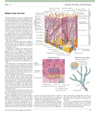
Plate 1-2                                                                             Anatomy, Physiology, and Embryology

                                                                        Free nerve endings  Meissner corpuscle  Stratum corneum
                                                                         Hair shaft           Pore of sweat gland  Stratum lucidum
                                                                 Melanocyte
        NORMAL SKIN ANATOMY                         Arrector muscle of hair                                        Stratum
                                                                                                                   granulosum
                                                    Sebaceous gland                                                               Epidermis
                                                     Cuticle                                                       Stratum spinosum
        The human skin, taken collectively, is the largest organ                                                   Stratum basale
        in the human body. On average, it weighs between 4   Internal
                                                     sheath
        and 5 kg. It is vitally important to life. The skin is made
        up of three distinct layers: the epidermis, the dermis,   External                                        Dermal papilla
        and  the  subcutaneous  tissue;  some  anatomists  do  not   Hair follicle  sheath                        (of papillary layer)
        include the subcutaneous tissue as part of the skin and   Glassy
        classify it separately as the hypodermis. Each of these   membrane
        layers plays a pivotal role in the execution of day-to-day                                                                Dermis
        functions  of  the  skin.  The  skin’s  main  function  is  to   Connective
        protect the interior of the body from the exterior envi-  tissue layer                                    Reticular layer
        ronment. It performs this role in many fashions: It acts   Hair cuticle
        as  a  semipermeable  barrier  to  both  hydrophilic  and
        hydrophobic substances; it is the first line of immuno-
        logical  defense  against  invading  microbes;  it  contains   Sweat gland
        many components of the adaptive and innate immune   Hair matrix
        system; and it has many physiological roles, including
        metabolism of vitamin D.                     Papilla of                                                                   Subcutaneous tissue
          The majority of the epidermis is made up of kerati-
        nocytes.  It  also  contains  melanocytes,  Langerhans     hair follicle
        cells, and Merkel cells. The epidermis is avascular and   Pacinian corpuscle
        receives its nutrition from the superficial vascular plexus   Artery
        of the papillary dermis.                                     Vein
          Melanocytes  are  derived  from  neural  crest  and
        are  responsible  for  producing  the  melanin  family  of     Sensory nerves                                  Subcutaneous
        pigments,  which  are  packaged  in  melanosomes.                    Elastic fibers                            artery and vein
        Melanocytes are found in equal density in all humans,        Skin ligaments (retinacula cutis)     Cutaneous nerve
        but darker-skinned individuals have a higher density of
        melanosomes than those with lighter skin. This is the                      Motor (autonomic) nerve
        reason for color variation among humans. Eumelanin,
        the predominant type of melanin protein, is responsible       Detail of Merkel disc                  Detail of free nerve ending
        for  brown  and  black  pigmentation.  Pheomelanin  is  a                                             Basement membrane
        unique variant of melanin that is found in humans with                                                    Axon terminal
        red hair.                                 Basal
          The  skin  is  found  in  continuity  with  the  epithelial   epithelial                                 Mitochondrion
        lining of the digestive tract, including the oral mucosa   cells                                           Schwann cell
        and the anal mucosa. Distinct transition zones are seen
        at these interfaces. The skin also abuts the conjunctival
        mucosa of the globe and the mucosa of the nasal pas-
        sages. The skin and its neighboring epithelial compo-  Cytoplasmic                             Cross section
        nents supply the human body with a continuous barrier   protrusion
        to protect it from the external world.
          Many appendageal structures are present throughout
        the  skin.  The  major  ones  are  the  hair  follicles,  their
        associated  sebaceous  glands,  and  the  eccrine  glands.   Mitochondria
        Most of the skin is hair bearing. Fine vellus hairs make
        up  the  preponderance  of  the  skin’s  hair  production.
        Terminal hairs are much thicker and are found on the
        scalp, eyebrows, and eyelashes; in the axilla and groin
        areas; and in the beard region in men. Glabrous skin,   Desmosomes                 Schwann cell
        which is devoid of hair follicles, includes the vermilion   Expanded axon terminal                     Axon
        border of the lips, the palms, the soles, the glans penis,                    Merkel cell
        and the labia minora.                                                        Lobulated nucleus
          Human skin varies in thickness. It is thickest on the                  Granulated vesicles      Schwann cells
        back, and the thinnest areas are found on the eyelids and
        the scrotum. Regardless of thickness, all skin possesses
        the same immunological function and barrier activity.
          Various appendageal structures are found in higher   The other main sweat glands of the skin, the apocrine   that  the  fingernails  grow  slightly  faster  than  the  toe-
        densities  in  certain  regions  of  the  skin.  Sebaceous   glands, are found almost exclusively in the axillae and   nails. The average thumbnail takes 6 months to replace
        glands  are  located  predominantly  on  the  face,  upper   the groin. The apocrine glands, like sebaceous glands,   itself,  whereas  the  average  great  toenail  takes  8  to
        chest, and back. These glands play an instrumental role   are found only in conjunction with hair follicles.  12 months.
        in the pathomechanism of acne vulgaris. Because seba-  Nails are composed of specialized keratin proteins.   Skin is also an important means of communication
        ceous  glands  are  attached  to  hair  follicles,  they  are   These keratins make a hard nail plate that is believed   with  other  humans.  The  sense  of  touch  is  mediated
        found only on hair-bearing skin. Eccrine sweat glands,   to be important for protection, grasp, and defense. Fin-  through  specialized  receptors  within  the  skin.  One
        on the other hand, are found ubiquitously. The highest   gernails and toenails are made of the same keratin struc-  cannot underestimate the importance of this function
        densities of eccrine glands are on the palms and soles.   ture  and  in  the  same  manner.  The  only  difference  is   in the formation of human relationships.


        THE NETTER COLLECTION OF MEDICAL ILLUSTRATIONS                                                                            3