
Page 19 - The Netter Collection of Medical Illustrations - Integumentary System_ Volume 4 ( PDFDrive )
P. 19
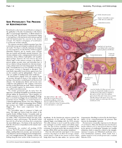
Plate 1-4                                                                             Anatomy, Physiology, and Embryology


                                                                                                               Bricks (keratinocytes)
                                                                                                               Mortar (intercellular space
                                                                                                               of the stratum corneum)
                                                     Cornified layer
        SKIN PHYSIOLOGY: THE PROCESS                                                                           Corneodesmosomes
        OF KERATINIZATION


        Keratinization, also known as cornification, is unique to
        the epithelium of the skin. Keratinization of the human
        skin is of paramount importance; it allows humans to
        live on dry land. The process of keratinization begins
        in the basal layer of the epidermis and continues upward
        until  full  keratinization  has  occurred  in  the  stratum   Granular  layer
        corneum. The function and purpose of keratinization
        is to form the stratum corneum.
          The stratum corneum is a highly organized layer that
        is relatively strong and resistant to physical and chemi-                                             Cornified cell envelope
        cal insults. This layer is critically important in keeping   Spinous                                  cross linked with ceramides
        out microorganisms; it is the first line of defense against   layer                                   replaces plasma membrane
        ultraviolet  radiation;  and  it  contains  many  enzymes
        that can degrade and detoxify external chemicals. The                                                              Filaments
        stratum corneum is also a semipermeable structure that                                        Corneodesmosome       of keratin
        selectively  allows  different  hydrophilic  and  lipophilic
        agents passage. However, the most obvious and most
        studied aspect of the stratum corneum is its ability to                                  LM
        protect against excessive water and electrolyte loss. It                                                          Corneocyte
        acts as a barrier to keep chemicals out, but more impor-  Basal layer
        tantly, it keeps water and electrolytes inside the human                             SG cell    LB
        body. Transepidermal water loss (TEWL) increases as                                                         LB
        the  stratum  corneum  is  damaged  or  disrupted.  The
        main lipids responsible for protection against water loss                                                       LB
        are the ceramides and the sphingolipids. These mole-
        cules are capable of binding many water molecules.
          As  keratinocytes  migrate  from  the  stratum  basale
        and journey through the layers of the epidermis, they   Dermis                          Keratohyalin
        undergo characteristic morphological and biochemical                                    granules
        changes.  The  keratinocytes  flatten  and  become  more
        compacted and polyhedral. The resulting corneocytes                                              Golgi
        become stacked, like bricks in a wall. These corneocytes                                         apparatus
        are  still  bonded  together  by  desmosomes,  which  are
        now called corneodesmosomes.                                                                  Lamellar bodies (LB) that are seen today
          The  stratum  granulosum  gets  its  name  from  the                                        as part of a branched tubular structure
        appearance of multiple basophilic keratohyalin granules                                       like the trans-Golgi network migrate to
        present  within  the  keratinocytes.  These  granules  are                                    the surface of the cell of the stratum
        largely composed of the protein profilaggrin. Profilag-  The dashed lines (                )  granulosum (SG) and release their content
        grin is converted into filaggrin by an intercellular endo-  show the tortuous intercellular   into the intercellular space (ICS). The
        proteinase enzyme. Filaggrin is so named because it is   penetration pathway within the       released lipids are rearranged into lamellar
        a filament-aggregating protein. Over time, filaggrin is   stratum corneum taken by water-     membrane (LM)
        broken down into natural moisturizing factor (NMF)   soluble substances when the permeability
        and  urocanic  acid.  NMF  is  a  breakdown  product  of   of the skin barrier is activated
        filaggrin  that  slows  water  evaporation  from  the
        corneocytes.
          The  intercellular  space  is  composed  of  lipids  and
        water.  The  lipids  are  derived  from  the  release  of  the
        lamellar  bodies  (Odland  bodies).  Ceramides  make  up
        the overwhelming majority of the contents of the lamel-
        lar bodies. Other components include free fatty acids,   membrane.  As  the  keratinocyte  migrates  upward,  the   desquamation. Shedding is achieved by the final degra-
        cholesterol esters, and proteases. The lamellar bodies   cell  membrane  is  lost,  and  the  ceramides  that  are   dation  of  the  corneodesmosomes  by  proteases  that
        fuse with the cell surface and release their contents into   released  begin  cross-linking  with  the  CCE  proteins.   destroy the desmoglein-1 protein.
        the intercellular space. The fusion of the lamellar body   The cells continue to move toward the surface of the   Keratinization is especially important in the diseases
        with the cell surface is dependent on the enzyme trans-  skin and begin to lose their nucleus and cellular organ-  of cornification. Many skin diseases have been found to
        glutaminase I.                            elles. The loss of these organelles is mediated by the   involve defects in one or more proteins that are critical
          Concurrently.  the  cornified  cell  envelope  (CCE)   activation of certain proteases that can quickly degrade   in the process of cornification. Examples are lamellar
        develops. The CCE proteins envoplakin, loricrin, peri-  protein, DNA, RNA, and the nuclear membrane.  ichthyosis, which is caused by a defect in the transglu-
        plakin, small proline-rich proteins, and involucrin are   Once the cells reach the outer layers of the stratum   taminase  I  enzyme,  and  Vohwinkel’s  syndrome  (kera-
        cross-linked in various arrangements by transglutamin-  corneum, they begin to be shed. On average, a kerati-  toma  hereditarium  mutilans),  which  results  from  a
        ase I and transglutaminase III, forming a sturdy scaf-  nocyte spends 2 weeks in the stratum corneum before   genetic mutation in the loricrin protein and a resultant
        folding along the inner surface of the keratinocyte cell   being  shed  from  the  skin  surface  in  a  process  called   defective CCE.


        THE NETTER COLLECTION OF MEDICAL ILLUSTRATIONS                                                                            5