
Page 253 - The Netter Collection of Medical Illustrations - Integumentary System_ Volume 4 ( PDFDrive )
P. 253
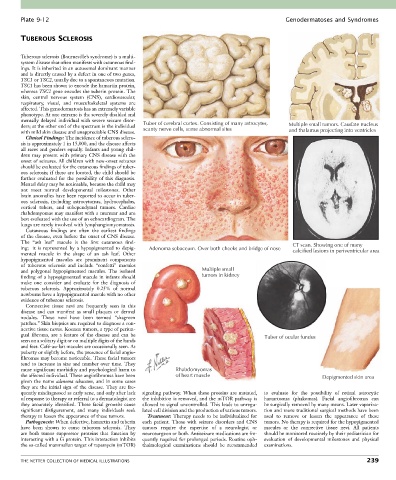
Plate 9-12                                                                                  Genodermatoses and Syndromes


        TUBEROUS SCLEROSIS


        Tuberous sclerosis (Bourneville’s syndrome) is a multi-
        system disease that often manifests with cutaneous find-
        ings. It is inherited in an autosomal dominant manner
        and is directly caused by a defect in one of two genes,
        TSC1 or TSC2, usually due to a spontaneous mutation.
        TSC1 has been shown to encode the hamartin protein,
        whereas TSC2 gene encodes the tuberin protein. The
        skin,  central  nervous  system  (CNS),  cardiovascular,
        respiratory,  visual,  and  musculoskeletal  systems  are
        affected. This genodermatosis has an extremely variable
        phenotype. At one extreme is the severely disabled and
        mentally delayed individual with severe seizure disor-  Tuber of cerebral cortex. Consisting of many astrocytes,
        ders; at the other end of the spectrum is the individual   scanty nerve cells, some abnormal sites  Multiple small tumors. Caudate nucleus
        with mild skin disease and unappreciable CNS disease.                                        and thalamus projecting into ventricles
          Clinical Findings: The incidence of tuberous sclero-
        sis is approximately 1 in 15,000, and the disease affects
        all races and genders equally. Infants and young chil-
        dren may present with primary CNS disease with the
        onset of seizures. All children with new-onset seizures
        should be evaluated for the cutaneous findings of tuber-
        ous sclerosis; if these are located, the child should be
        further  evaluated  for  the  possibility  of  this  diagnosis.
        Mental delay may be noticeable, because the child may
        not  meet  normal  developmental  milestones.  Other
        brain anomalies have been reported to occur in tuber-
        ous  sclerosis,  including  astrocytomas,  hydrocephalus,
        cortical  tubers,  and  subependymal  tumors.  Cardiac
        rhabdomyomas may manifest with a murmur and are
        best evaluated with the use of an echocardiogram. The
        lungs are rarely involved with lymphangiomyomatosis.
          Cutaneous  findings  are  often  the  earliest  findings
        of the disease, even before the onset of CNS disease.
        The  “ash  leaf”  macule  is  the  first  cutaneous  find-                                    CT scan. Showing one of many
        ing;  it  is  represented  by  a  hypopigmented  to  depig-  Adenoma sebaceum. Over both cheeks and bridge of nose  calcified lesions in periventricular area
        mented  macule  in  the  shape  of  an  ash  leaf.  Other
        hypopigmented  macules  are  prominent  components
        of  tuberous  sclerosis  and  include  “confetti”  macules
        and  polygonal  hypopigmented  macules.  The  isolated         Multiple small
        finding of a hypopigmented macule in infants should            tumors in kidney
        make  one  consider  and  evaluate  for  the  diagnosis  of
        tuberous  sclerosis.  Approximately  0.25%  of  normal
        newborns have a hypopigmented macule with no other
        evidence of tuberous sclerosis.
          Connective  tissue  nevi  are  frequently  seen  in  this
        disease  and  can  manifest  as  small  plaques  or  dermal
        nodules.  These  nevi  have  been  termed  “shagreen
        patches.” Skin biopsies are required to diagnose a con-
        nective tissue nevus. Koenen tumors, a type of periun-
        gual  fibroma,  are  a  feature  of  the  disease  and  can  be                     Tuber of ocular fundus
        seen on a solitary digit or on multiple digits of the hands
        and feet. Café-au-lait macules are occasionally seen. At
        puberty or slightly before, the presence of facial angio-
        fibromas may become noticeable. These facial tumors
        tend to increase in size and number over time. They
        cause significant morbidity and psychological harm to   Rhabdomyomas
        the affected individual. These angiofibromas have been   of heart muscle                                Depigmented skin area
        given  the  name  adenoma  sebaceum,  and  in  some  cases
        they  are  the  initial  sign  of  the  disease.  They  are  fre-
        quently misdiagnosed as early acne, and only after lack   signaling  pathway.  When  these  proteins  are  mutated,   to  evaluate  for  the  possibility  of  retinal  astrocytic
        of response to therapy or referral to a dermatologist are   the inhibition is removed, and the mTOR pathway is   hamartomas  (phakomas).  Facial  angiofibromas  can
        they accurately identified. These facial growths cause   allowed to signal uncontrolled. This leads to unregu-  be surgically removed by many means. Laser vaporiza-
        significant  disfigurement,  and  many  individuals  seek   lated cell division and the production of various tumors.  tion and more traditional surgical methods have been
        therapy to lessen the appearance of these tumors.  Treatment: Therapy needs to be individualized for   used  to  remove  or  lessen  the  appearance  of  these
          Pathogenesis: When defective, hamartin and tuberin   each  patient.  Those  with  seizure  disorders  and  CNS   tumors. No therapy is required for the hypopigmented
        have  been  shown  to  cause  tuberous  sclerosis.  They     tumors  require  the  expertise  of  a  neurologist  or     macules  or  the  connective  tissue  nevi.  All  patients
        are  both  tumor  suppressor  proteins  that  function  by   neurosurgeon or both. Antiseizure medications are fre-  should be monitored routinely by their pediatrician for
        interacting with a G protein. This interaction inhibits   quently required for prolonged periods. Routine oph-  evaluation  of  developmental  milestones  and  physical
        the so-called mammalian target of rapamycin (mTOR)   thalmological  examinations  should  be  recommended     examinations.


        THE NETTER COLLECTION OF MEDICAL ILLUSTRATIONS                                                                          239