
Page 250 - The Netter Collection of Medical Illustrations - Integumentary System_ Volume 4 ( PDFDrive )
P. 250
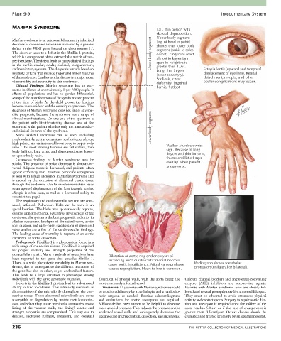
Plate 9-9                                                                                             Integumentary System


       MARFAN SYNDROME                                                       Tall, thin person with
                                                                             skeletal disproportion.
       Marfan syndrome is an autosomal dominantly inherited                  Upper body segment
                                                                             (top of head to pubis)
       disorder of connective tissue that is caused by a genetic             shorter than lower body
       defect in the FBN1 gene located on chromosome 15.                     segment (pubis to soles
       The disorder leads to a defect in the fibrillin-1 protein,          Upper body segment  of feet). Fingertips reach
       which is a component of the extracellular matrix of con-              almost to knees (arm
       nective tissue. The defect leads to many clinical findings            span-to-height ratio
       in  the  cardiovascular,  ocular,  skeletal,  integumentary,          greater than 1.05).
       and respiratory systems. The diagnosis is made based on               Long, thin fingers     Ectopia lentis (upward and temporal
       multiple criteria that include major and minor features               (arachnodactyly).      displacement of eye lens). Retinal
       of the syndrome. Cardiovascular disease is a major cause              Scoliosis, chest       detachment, myopia, and other
       of morbidity and mortality in this syndrome.                          deformity, inguinal    ocular complications may occur.
         Clinical  Findings:  Marfan  syndrome  has  an  esti-               hernia, flatfoot
       mated incidence of approximately 1 per 7500 people. It
       affects all populations and has no gender differential.
       Many of the manifestations of the syndrome are present
       at the time of birth. As the child grows, the findings
       become more evident and the severity may worsen. The
       diagnosis of Marfan syndrome does not imply any spe-
       cific  prognosis,  because  the  syndrome  has  a  range  of
       clinical manifestations. On one end of the spectrum is
       the  patient  with  life-threatening  disease,  and  at  the
       other end is the patient who has only the musculoskel-              Lower body segment
       etal clinical features of the syndrome.
         Many  skeletal  anomalies  can  be  seen,  including
       arachnodactyly, pectus excavatum, scoliosis, pes planus,
       high palate, and an increased lower body to upper body
       ratio. The most striking features are tall stature, thin                   Walker-Murdoch wrist
       body habitus, long arms, and disproportionate lower-                       sign. Because of long
       to-upper body ratio.                                                       fingers and thin forearm,
         Cutaneous  findings  of  Marfan  syndrome  may  be                       thumb and little finger
       subtle. The presence of striae distensae is almost uni-                    overlap when patient
       versal. Adipose tissue is decreased, and patients often                    grasps wrist.
       appear extremely thin. Elastosis perforans serpiginosa
       is seen with a high incidence in Marfan syndrome and
       is  caused  by  the  extrusion  of  abnormal  elastic  tissue
       through the epidermis. Ocular involvement often leads
       to an upward displacement of the lens (ectopia lentis).
       Myopia is often seen, as well as a decreased ability to
       constrict the pupil.
         The respiratory and cardiovascular systems are com-
       monly  affected.  Pulmonary  blebs  can  be  seen  in  an
       apical location. The blebs may spontaneously rupture,
       causing a pneumothorax. Severity of involvement of the
       cardiovascular system is the best prognostic indicator in
       Marfan syndrome. Prolapse of the mitral valve, aortic
       root dilation, and early-onset calcification of the mitral
       valve  anulus  are  a  few  of  the  cardiovascular  findings.
       The leading cause of mortality is rupture of an aortic
       aneurysm or aortic dissection.
         Pathogenesis: Fibrillin-1 is a glycoprotein found in a
       wide range of connective tissues. Fibrillin-1 is required
       for  proper  elasticity  and  strength  properties  of  the
       extracellular matrix. Many hundreds of mutations have   Dilatation of aortic ring and aneurysm of
       been  reported  in  the  gene  that  encodes  fibrillin-1.   ascending aorta due to cystic medial necrosis
       There is a wide phenotypic variability in Marfan syn-  cause aortic insufficiency. Mitral valve prolapse  Radiograph shows acetabular
       drome, due in some part to the different mutations of   causes regurgitation. Heart failure is common.  protrusion (unilateral or bilateral).
       the gene but also to other, as yet undescribed factors.
       This  leads  to  a  large  variation  in  phenotype  among
       individuals with the same genotypic mutation.  dissection  of  arterial  walls,  with  the  aorta  being  the   Calcium channel blockers and angiotensin-converting
         Defects in the fibrillin-1 protein lead to a decreased   most commonly affected vessel.  enzyme  (ACE)  inhibitors  are  second-line  agents.
       ability to bind to calcium. This ultimately manifests as   Treatment: All patients with Marfan syndrome should   Patients  with  Marfan  syndrome  who  are  closely  fol-
       abnormalities of the microfibrils throughout the con-  be monitored directly by a cardiologist and a cardiotho-  lowed and treated promptly may live a normal life span.
       nective  tissue.  These  abnormal  microfibrils  are  more   racic  surgeon  as  needed.  Routine  echocardiograms     They  must  be  educated  to  avoid  strenuous  physical
       susceptible  to  degradation  by  matrix  metalloprotein-  and  evaluations  for  aortic  aneurysms  are  required.   activity and contact sports. Surgery to repair aortic dila-
       ases, and when they occur within the connective tissue   β-Blockade has been shown to be helpful to decrease   tion and aneurysm is required once the caliber of the
       lining  of  the  vascular  walls,  the  lining’s  elastic  and   mean arterial pressure. This reduces the pressure on the   aorta  reaches  5.0 cm  or  if  the  rate  of  enlargement  is
       strength properties are compromised. This may lead to   weakened vessel walls and subsequently decreases the   greater  than  0.5 cm/year.  Ocular  disease  should  be
       dilation,  increased  stiffness,  aneurysm,  and  eventual   likelihood of arterial dilation, dissection, and aneurysms.   evaluated and treated promptly by an ophthalmologist.

       236                                                                                   THE NETTER COLLECTION OF MEDICAL ILLUSTRATIONS