
Page 251 - The Netter Collection of Medical Illustrations - Integumentary System_ Volume 4 ( PDFDrive )
P. 251
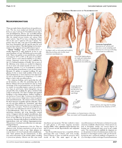
Plate 9-10                                                                                  Genodermatoses and Syndromes

                                                                       CUTANEOUS MANIFESTATIONS OF NEUROFIBROMATOSIS


        NEUROFIBROMATOSIS


        There are eight distinct clinical forms of neurofibroma-
        tosis.  The  two  most  studied  and  clinically  important
        forms are type I and type II. Neurofibromatosis type I
        (von  Recklinghausen  disease)  and  neurofibromatosis
        type II are autosomal dominant disorders involving the
        skin,  the  central  nervous  system,  and  various  other
        organ systems. Type II has many overlapping features
        that are also seen in patients with type I disease. The
        genetic bases for type I and type II neurofibromatosis
        have been determined, and the specific gene for each                                                Verrucous hyperplasia.
        type has been isolated. The skin findings can be instru-                                            Maceration of velvety-soft
        mental in the diagnosis of neurofibromatosis type I.                                                skin may cause weeping and
          Clinical  Findings:  Type  I  neurofibromatosis  is                                               infection in crevices overlying
        usually  diagnosed  in  early  childhood.  It  has  an  esti-  Multiple café-au-lait spots and nodules  a plexiform neurofibroma.
                                                    (fibroma molluscum) are the most
        mated incidence of 1 per 3000 births and occurs world-  common manifestations.
        wide. There is no gender or race predilection, and type
        I accounts for 85% to 90% of all cases of neurofibro-                        Localized elephantiasis
        matosis. There is wide clinical variability in neurofibro-                   of thigh with redundant
        matosis.  Diagnostic  criteria  have  been  established  by                  skin folds overlying a
        the U.S. National Institutes of Health. Two or more of                       plexiform neurofibroma.
        the  following  seven  criteria  are  needed  for  diagnosis:
        (1) six or more café-au-lait macules (≥5 mm in size in
        prepuberty patients; >1.5 cm in postpuberty patients);
        (2) one plexiform neurofibroma or two or more neuro-
        fibromas;  (3)  axillary  or  inguinal  freckling;  (4)  optic
        glioma; (5) two or more Lisch nodules of the iris; (6)
        sphenoid dysplasia or other distinctive bone abnormal-
        ity, such as pseudarthrosis of a long bone; or (7) a first-
        degree relative with neurofibromatosis.
          The cutaneous findings, and in particular the café-
        au-lait  macules,  are  often  the  presenting  sign  of  the
        disease. Solitary café-au-lait macules are seen in a large
        percentage of the normal population, and the diagnos-                    Large plexiform
        tic criteria for neurofibromatosis require the presence                  neurofibroma
        of at least six such lesions. Spinal dysraphism may be                   localized to one
        present if the skin overlying the spine is involved with                 side of trunk
        a café-au-lait macule. The onset of axillary and inguinal                and thigh
        freckling is often during puberty. Axillary freckling is
        also known as Crowe’s sign. Cutaneous neurofibromas
        are the most common benign tumor found in patients
        with neurofibromatosis. The tumors tend to be plenti-
        ful and to increase in number and size with time. They
        are soft and often exhibit the “buttonhole” sign when
        compressed. These tumors may have an overlying pink                                           Dense axillary and inguinal freckling
        to light violet coloration. Plexiform neurofibromas are                                       is rarely found in the absence of NF1.
        large dermal and subcutaneous tumors specific to type
        I  neurofibromatosis.  They  can  cause  compression  of
        underlying  structures  and  wrap  themselves  around
        nerves. Compared with the typical neurofibroma, they                Lisch nodules are hamartomas of the iris.
        are firm and larger and have an ill-defined border. Both            They are raised and frequently pigmented.
        forms of neurofibromas can produce varying amounts
        of pruritus. Patients with plexiform neurofibromas have
        hypertrichosis  with  and  without  hyperpigmentation.
        The presence of multiple neurofibromas can cause psy-  disturbance and proptosis. The best method to detect   neurofibromatosis is the formation of bilateral acoustic
        chological disease.                       an  optic  glioma  is  with  brain  magnetic  resonance   neuromas (vestibular schwannomas). These tumors can
          Lisch nodules are hamartomas of the iris. They are   imaging (MRI). Other ophthalmological findings that   lead  to  headaches,  vertigo,  and  various  degrees  of
        observed under slit-lamp examination and can be seen   may  be  present  include  hypertelorism  and  congenital   hearing  loss.  Schwannomas  may  occur  in  any  cranial
        by  approximately  6  years  of  age.  Optic  gliomas  are     glaucoma.            nerve. The criteria used to establish the diagnosis are
        seen in about 1 of every 8 patients with neurofibroma-  Type II neurofibromatosis has a completely different   (1) the presence of bilateral schwannomas; (2) the com-
        tosis.  Optic  gliomas  may  be  asymptomatic,  or  they     phenotype  than  type  I  disease,  with  some  overlap.   bination of a first-degree relative with type II neurofi-
        may cause compression of the pituitary gland, resulting   Onset of disease is often not until the second or third   bromatosis and a unilateral vestibular schwannoma; or
        in  precocious  puberty.  Gliomas  can  also  cause  visual   decade  of  life.  The  main  aspect  of  type  II   (3) a first-degree relative with type II neurofibromatosis


        THE NETTER COLLECTION OF MEDICAL ILLUSTRATIONS                                                                          237