
Page 252 - The Netter Collection of Medical Illustrations - Integumentary System_ Volume 4 ( PDFDrive )
P. 252
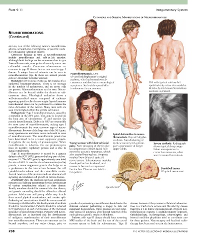
Plate 9-11                                                                                            Integumentary System

                                                                 CUTANEOUS AND SKELETAL MANIFESTATIONS OF NEUROFIBROMATOSIS


       NEUROFIBROMATOSIS
       (Continued)


       and  any  two  of  the  following  tumors:  neurofibroma,
       glioma, schwannoma, meningioma, or juvenile poste-
       rior subcapsular lenticular opacity.
         Cutaneous  findings  in  type  II  neurofibromatosis
       include  neurofibromas  and  café-au-lait  macules.
       Although both findings are less numerous than in type
       I neurofibromatosis, most patients have only one or two
       café-au-lait  macules.  Cutaneous  schwannomas  are
       common in type II disease but are not seen in type I
       disease.  A  unique  form  of  cataracts  can  be  seen  in
       neurofibromatosis  type  II;  these  are  termed  juvenile   Neurofibromatosis. One
       posterior subcapsular lenticular cataracts.  of von Recklinghausen’s original
         Histology: Skin biopsies of café-au-lait macules show   patients, who had extensive sub-
       epidermal  hyperpigmentation.  There  is  no  increase     cutaneous nodules but no neurological  Girl with typical café-au-lait
                                                                                                        spots but only a few skin nodules.
       in  the  number  of  melanocytes,  and  no  nevus  cells     symptoms. Such wide-spread skin     Relatively mild neurofibromatous
       are present. Macromelanosomes can be seen. Neuro-  involvement is uncommon.                      scoliosis is present.
       fibromas  can  be  located  within  the  dermis  or  sub-
       cutaneous  tissue.  Histological  evaluation  shows  a
       well-circumscribed  tumor  composed  of  uniform-
       appearing spindle cells of nerve origin. Special immune
       histochemical stains can be performed to confirm the
       nerve  derivation  of  the  tumors.  Many  mast  cells  are
       seen intermingled within the spindle cell tumor.
         Pathogenesis: Type I neurofibromatosis is caused by
       a mutation in the NF1 gene. This gene is located on
       the  long  arm  of  chromosome  17  and  encodes  the
       protein neurofibromin. Defects in NF1 are responsible
       for  most  cases  of  neurofibromatosis,  making  type  I
       neurofibromatosis  the  most  common  type  of  neuro-
       fibromatosis. Because of the large size of the NF1 gene,
       many spontaneous mutations occur and result in cases                    Spinal deformities in neuro-
       of  neurofibromatosis.  The  neurofibromin  protein  has                fibromatosis. Boy with kypho-
       been  determined  to  be  a  tumor  suppressor  protein.                scoliosis. Foreshortening of
       It  regulates  the  ras  family  of  protooncogene.  When   Young woman with bilateral facial  trunk secondary to kyphosis  Severe scoliosis. Radiograph
       neurofibromin  is  defective,  the  ras  protooncogene   palsy. Note drooping of cheeks due  gives appearance of longer  shows typical sharp angu-
       loses  its  negative  regulatory  protein  and  is  able  to     to compression of both facial (VII)  upper limbs.  lation unresponsive to
       signal continuously.                      nerves by acoustic neuromas, which                        corrective measures, often
         Type  II  neurofibromatosis  is  caused  by  a  genetic   also caused hearing loss. Proptosis
       defect in the SCH (NF2) gene on the long arm of chro-  resulted from bilateral optic (II)           seen in neurofibromatosis.
       mosome 22. The NF2 gene is approximately one third   nerve tumors. Subcutaneous nodules
       the size of NF1. It encodes the schwannomin (merlin)   developed on her forehead, and
       protein,  a  tumor  suppressor  protein  that  helps  act  as     masses in her neck compressed
       a  go-between  in  the  interactions  between  the  cell   the trachea. Disease was fatal in             Dumbbell tumor
       cytoskeleton/membrane  and  the  extracellular  matrix.   this patient.                                  Of spinal nerve root
       Loss of function of the protein results in abnormal cell
       signaling and unabated cell growth in various tissues.
         Treatment: Once the diagnosis has been established,
       patients need lifelong monitoring for the development
       of  various  complications  related  to  their  disease.           Spinal cord
       Family  members  should  be  screened  for  the  disease,
       and  genetic  counseling  should  be  offered  to  affected
       patients.  Adolescents  and  young  adults  may  benefit
       from  annual  physical  examinations,  and  routine  oph-
       thalmological  examinations  should  be  recommended.
       Screening in childhood for the development of scoliosis   growth of a preexisting neurofibroma should make the   disease, because of the presence of bilateral schwanno-
       should be recommended. Patients should be screened   clinician  consider  performing  a  biopsy  to  rule  out   mas,  is  a  much  more  serious  and  life-altering  disease
       for hypertension at each visit because of the increased   malignant degeneration. Optic gliomas are best surgi-  than type I. The follow-up management of type II neu-
       incidence of pheochromocytoma. Patients with neuro-  cally excised if indicated, even though removal of the   rofibromatosis  requires  a  multidisciplinary  approach.
       fibromatosis  are  at  increased  risk  for  development     optic glioma typically results in blindness.  Ophthalmology,  otolaryngology,  neurosurgery,  and
       of  malignant  transformation  of  their  neurofibromas   Patients with type II disease should have screening   internal  medicine  physicians  need  to  coordinate  care
       into neurofibrosarcomas. These rare sarcomas can be   MRI  studies  of  the  brain  and  the  rest  of  the  central   for  these  patients.  Neurosurgery  and  localized  radio-
       located  anywhere,  and  any  major  change,  pain,  or   nervous  system  to  look  for  schwannomas.  Type  II   therapy have been used to treat the brain tumors.

       238                                                                                   THE NETTER COLLECTION OF MEDICAL ILLUSTRATIONS