
Page 249 - The Netter Collection of Medical Illustrations - Integumentary System_ Volume 4 ( PDFDrive )
P. 249
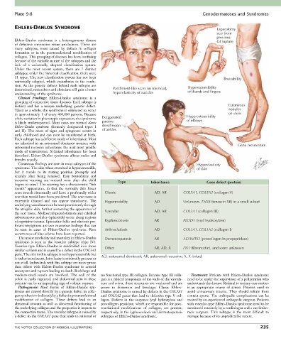
Plate 9-8                                                                                   Genodermatoses and Syndromes


        EHLERS-DANLOS SYNDROME                                                                             Laparotomy
                                                                                                           scar from
                                                                                                           previous
        Ehlers-Danlos  syndrome  is  a  heterogeneous  disease                                             GI rupture
        of  defective  connective  tissue  production.  There  are
        many  subtypes,  most  caused  by  defects  in  collagen
        formation  or  in  the  posttranslational  modification  of
        collagen. This grouping of diseases has been confusing
        because of the variable nature of the subtypes and the
        lack  of  a  universally  adopted  classification  system.
        Under  the  most  recent  system,  there  are  7  distinct
        subtypes; under the historical classification, there were
        11  types.  The  new  classification  system  has  not  been                                          Bruisability
        universally  adopted,  which  contributes  to  the  confu-
        sion.  As  the  genetic  defects  behind  each  subtype  are                        Hyperextensibility
        determined, researchers and clinicians will gain a better   Parchment-like scars on forehead,  of thumb and fingers
        understanding of the syndrome.                  hyperelasticity of auricles
          Clinical  Findings:  Ehlers-Danlos  syndrome  is  a
        grouping of connective tissue diseases. Each subtype is
        distinct  and  has  a  unique  underlying  genetic  defect.                                             Cutaneous
        Taken as a whole, the syndrome is estimated to occur                                                    nodules
        in approximately 1 of every 400,000 persons. Because                                Hyperextensibility  on shins
        of the variation in phenotypic expression, the syndrome   Exaggerated               of elbows
        is  likely  underreported.  Most  cases  are  termed  classic   passive
        Ehlers-Danlos  syndrome  (formerly  designated  types  I   dorsiflexion
        and  II).  The  onset  of  signs  and  symptoms  occurs  in   of ankles
        early  childhood  and  can  even  be  manifested  at  birth.
        Each subtype has a different mode of inheritance. Most
        are inherited in an autosomal dominant manner, with                                                          Genu recurvatum
        autosomal recessive inheritance the next most prolific
        mode  of  transmission.  X-linked  inheritance  has  been
        described.  Ehlers-Danlos  syndrome  affects  males  and
        females equally.
          Cutaneous findings are seen in most subtypes of the                                    Hyperelasticity
        syndrome. The skin when stretched is hyperextensible,                                    of skin
        but  it  recoils  to  its  resting  position  promptly  and
        entirely  after  being  released.  Easy  bruisability  and
        excessive  scarring  are  noticed  soon  after  the  child   Type  Inheritance                Gene defect (protein)
        begins to crawl. The scarring has a characteristic “fish
        mouth”  appearance,  in  that  the  normally  thin  linear
        scars stretch abnormally and leave a profoundly wider   Classic     AD, AR        COL5A1, COL5A2 (collagen V)
        scar than would have been predicted. The scar tissue is
        extremely  thinned  and  can  appear  translucent.  The   Hypermobility  AD       Unknown, TNXB (tenascin XB) in a small subset
        underlying vasculature can be seen prominently through
        the atrophic skin, further worsening the appearance of   Vascular   AD, AR        COL3A1 (collagen III)
        the scar tissue. Molluscoid pseudotumors and calcified
        subcutaneous nodules (spheroids) occur along regions
        of repetitive trauma. Epicanthic folds and elastosis per-  Kyphoscoliosis   AR    PLOD1 (lysyl hydroxylase)
        forans serpiginosa are two cutaneous findings that can
        be  seen  in  cases  of  Ehlers-Danlos  syndrome.  Rare   Arthrochalasis  AD      COL1A1, COL1A2 (collagen I)
        occurrences of blue sclerae have been reported.
          The major morbidity and mortality in Ehlers-Danlos   Dermatosparaxis  AR        ADAMTS2 (procollagen In-propeptidase)
        syndrome  is  seen  in  the  vascular  subtype  (type  IV).
        Vascular-type  Ehlers-Danlos  is  subdivided  into  three   Other   AR, AD, X     FN1 (fibronectin), and some unknown
        similar variants and is caused by a defect in the COL3A1
        gene. The skin in this subtype is not hyperextensible but   AD, autosomal dominant; AR, autosomal recessive; X, X-linked
        is rather translucent. Joint laxity is minimally present or
        not at all. Individuals with this subtype are more prone
        than  others  with  Ehlers-Danlos  syndrome  to  arterial
        aneurysms and rupture leading to death. Both large and
        medium-sized  vessels  are  involved.  The  wall  of  the   no functional type III collagen. Because type III colla-  Treatment:  Patients  with  Ehlers-Danlos  syndrome
        colon is easily ruptured, and abdominal pain in these   gen is a critical component of the walls of the vascula-  need to be under the supervision of a pediatrician who
        patients can be an impending sign of colonic rupture.  ture and colon, these structures are weakened and are   understands the disease. Referral to tertiary care centers
          Pathogenesis:  Most  forms  of  Ehlers-Danlos  syn-  prone  to  distention  and  breakage.  Classic  Ehlers-  is  an  appropriate  course  of  action.  Patients  need  to
        drome are caused directly by a genetic defect in colla-  Danlos syndrome is caused by defects in the COL5A1   avoid  unnecessary  trauma.  They  should  refrain  from
        gen synthesis or indirectly by a defect in posttranslational   and COL5A2 genes that lead to defective type V col-  contact  sports.  The  orthopedic  complications  can  be
        modification  of  collagen.  These  defects  lead  to  an   lagen.  Defects  in  the  enzymes  lysyl  hydroxylase  and   treated by an experienced orthopedic surgeon. Patients
        abnormal  amount  as  well  as  abnormal  functioning  of   procollagen peptidase, which are responsible for post-  with vascular-type Ehlers-Danlos syndrome need to be
        the underlying collagen and the properties it imparts to   translational  modifications  of  collagen,  are  present,   monitored routinely by a cardiologist and a cardiotho-
        the connective tissue. The vascular subtype is caused by   respectively, in the kyphoscoliosis and dermatosparaxis   racic  surgeon.  This  subtype  is  the  most  difficult  to
        a defect in the COL3A1 gene that leads to minimal or   subtypes of Ehlers-Danlos syndrome.  manage because of its unpredictable nature.


        THE NETTER COLLECTION OF MEDICAL ILLUSTRATIONS                                                                          235