
Page 194 - Clinical Application of Mechanical Ventilation
P. 194
160 Chapter 6
New York: Springer-Verlag. Used with permission. From Finucane, B. T., & Santora, A. H. (2003), Principles of airway management (3rd ed.).
In general, a size 7.5 or
8.0 ET tube should be used
for oral intubation of an adult
male. For an adult female, a
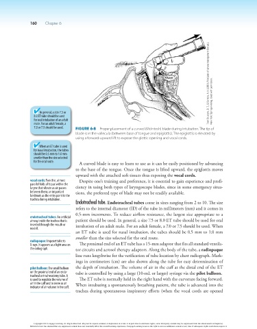
7.0 or 7.5 should be used. Figure 6-8 Proper placement of a curved (McIntosh) blade during intubation. The tip of
blade is in the vallecula (between base of tongue and epiglottis). The epiglottis is elevated by
using a forward-upward lift to expose the glottic opening and vocal cords.
When an ET tube is used
for nasal intubation, the tubes
should be 0.5 mm to 1.0 mm
smaller than the size selected
for the oral route. A curved blade is easy to learn to use as it can be easily positioned by advancing
to the base of the tongue. Once the tongue is lifted upward, the epiglottis moves
upward with the attached soft tissues thus exposing the vocal cords.
vocal cords: Two thin, almost Despite one’s training and preference, it is essential to gain experience and profi-
parallel folds of tissue within the
larynx that vibrate as air passes ciency in using both types of laryngoscope blades, since in some emergency situa-
between them; an important tions, the preferred type of blade may not be readily available.
landmark as the entry point to the
trachea during intubation.
Endotracheal tube. Endotracheal tubes come in sizes ranging from 2 to 10. The size
refers to the internal diameter (ID) of the tube in millimeters (mm) and it comes in
0.5-mm increments. To reduce airflow resistance, the largest size appropriate to a
endotracheal tubes: An artificial
airway inside the trachea that is patient should be used. In general, a size 7.5 or 8.0 ET tube should be used for oral
inserted through the mouth or intubation of an adult male. For an adult female, a 7.0 or 7.5 should be used. When
nostril.
an ET tube is used for nasal intubation, the tubes should be 0.5 mm to 1.0 mm
smaller than the size selected for the oral route.
radiopaque: Impenetrable to
X-rays. It appears as a light area on The proximal end of an ET tube has a 15-mm adaptor that fits all standard ventila-
the radiograph. tor circuits and aerosol therapy adaptors. Along the body of the tube, a radiopaque
line runs lengthwise for the verification of tube location by chest radiograph. Mark-
ings in centimeters (cm) are also shown along the tube for easy determination of
pilot balloon: The small balloon the depth of intubation. The volume of air in the cuff at the distal end of the ET
on the proximal end of an endo- tube is controlled by using a large (10-mL or larger) syringe via the pilot balloon.
tracheal or tracheostomy tube. It
is used to regulate the volume of The ET tube is normally held in the right hand with the curvature facing forward.
air in the cuff and to serve as an When intubating a spontaneously breathing patient, the tube is advanced into the
indicator of air volume in the cuff.
trachea during spontaneous inspiratory efforts (when the vocal cords are opened
Copyright 2013 Cengage Learning. All Rights Reserved. May not be copied, scanned, or duplicated, in whole or in part. Due to electronic rights, some third party content may be suppressed from the eBook and/or eChapter(s).
Editorial review has deemed that any suppressed content does not materially affect the overall learning experience. Cengage Learning reserves the right to remove additional content at any time if subsequent rights restrictions require it.