
Page 198 - Clinical Application of Mechanical Ventilation
P. 198
164 Chapter 6
TABLE 6-4 Procedure for Oral Intubation
1. Assemble and test supplies (e.g., check light source and ET tube cuff for air leak).
2. Lubricate the deflated cuff with a water-soluble lubricant.
3. Inform or explain procedure to patient.
4. Bag-mask ventilate and preoxygenate patient with 100% oxygen.
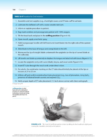
5. Tilt the head back and place in the sniffing position (Figure 6-10).
6. Open mouth, apply anesthetic spray.
7. Hold laryngoscope handle with left hand and insert blade into the right side of the opened
mouth.
8. Slide blade to the base of tongue and sweep blade to the left.
9. Maneuver the tip of straight blade underneath the epiglottis (or the tip of curved blade at
the vallecula).
10. Lift handle and blade up anteriorly to displace the tongue and attached soft tissues (Figure 6-11).
11. Locate the epiglottis (only with curve blade), larynx, and vocal cords (Figure 6-12).
12. Insert ET tube through the vocal cords under direct vision.
13. For adults, the centimeter marking on the ET tube should initially be placed at the lips or
incisors at 21 to 23 cm.
14. Inflate cuff and confirm endotracheal tube placement (e.g., loss of phonation, rising SpO ,
2
presence of bilateral breath sounds and expired CO ).
2
15. Verify proper depth of ET tube placement (1.5 inch above carina) with chest radiograph.
© Cengage Learning 2014
sniffing position: An ideal
head position for endotracheal
intubation. It is done by tilting the
forehead back slightly and moving
the mandible anteriorly to the
patient.
© Cengage Learning 2014
Figure 6-10 The head tilt (sniffing) position is done by tilting the forehead back slightly and
moving the mandible anteriorly to the patient.
Copyright 2013 Cengage Learning. All Rights Reserved. May not be copied, scanned, or duplicated, in whole or in part. Due to electronic rights, some third party content may be suppressed from the eBook and/or eChapter(s).
Editorial review has deemed that any suppressed content does not materially affect the overall learning experience. Cengage Learning reserves the right to remove additional content at any time if subsequent rights restrictions require it.