
Page 527 - Clinical Application of Mechanical Ventilation
P. 527
Critical Care Issues in Mechanical Ventilation 493
Treatment modalities for many critical conditions tend to target corrections of the
underlying causes (e.g., antibiotics for infection). At the present time, the best known
and most common management strategy for ALI and ARDS is supportive care for
oxygenation and ventilation. Studies on lung injury have identified risk factors and
have suggested that certain critical care interventions may influence the incidence of
lung injury. In the future, a well-designed screening tool or a lung injury predictive
model may help to reduce the incidence of ALI and ARDS (Litell et al., 2011).
Clinical Presentations
In the early stage of ARDS, the clinical signs may include tachypnea, tachycardia,
A combination of severe and mild hypoxemia. The patient’s oxygenation status worsens due to V/Q mis-
hypoxia, increased deadspace,
decreased lung compliance, match and intrapulmonary shunting. The PaO /F O ratio continues to decrease
2
I
2
and patient fatigue contrib-
utes to the development of as ARDS progresses. Severe hypoxia becomes evident with increasing deadspace
ventilatory failure. ventilation and decreasing lung compliance. When the patient cannot keep up with
the increasing work of breathing and oxygen demand, the PaCO begins to increase
2
and progresses to severe respiratory acidosis. Most patients develop diffuse alveolar
During the exudative
phase of ALI and ARDS, chest infiltrates and eventual respiratory failure within 48 hours of the onset of symptoms
radiographs reveal a progres- (Mortelliti et al., 2002).
sion from diffuse interstitial
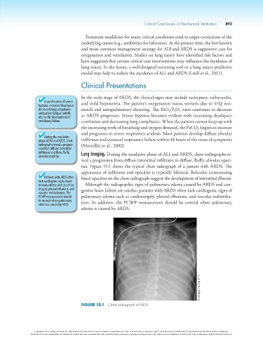
infiltrates to diffuse, fluffy, Lung Imaging. During the exudative phase of ALI and ARDS, chest radiographs re-
alveolar opacities.
veal a progression from diffuse interstitial infiltrates to diffuse, fluffy, alveolar opaci-
ties. Figure 15-1 shows the typical chest radiograph of a patient with ARDS. The
appearance of infiltrates and opacities is typically bilateral. Reticular (crisscrossing
Patients with ARDS often lines) opacities on the chest radiograph suggest the development of interstitial fibrosis.
lack cardiogenic signs of pul-
monary edema such as cardio- Although the radiographic signs of pulmonary edema caused by ARDS and con-
megaly, pleural effusions, and gestive heart failure are similar, patients with ARDS often lack cardiogenic signs of
vascular redistribution. The
PCWP measurement should pulmonary edema such as cardiomegaly, pleural effusions, and vascular redistribu-
be normal when pulmonary tion. In addition, the PCWP measurement should be normal when pulmonary
edema is casued by ARDS.
edema is caused by ARDS.
© Cengage Learning 2014
Figure 15-1 Chest radiograph of ARDS.
Copyright 2013 Cengage Learning. All Rights Reserved. May not be copied, scanned, or duplicated, in whole or in part. Due to electronic rights, some third party content may be suppressed from the eBook and/or eChapter(s).
Editorial review has deemed that any suppressed content does not materially affect the overall learning experience. Cengage Learning reserves the right to remove additional content at any time if subsequent rights restrictions require it.