
Page 532 - Clinical Application of Mechanical Ventilation
P. 532
498 Chapter 15
VAP range from 33 to 50%. The risk of VAP is highest immediately after intuba-
Risk factors for VAP tion and initiation of mechanical ventilation. For the first 5 days, the incidence of
include long duration of me-
chanical ventilation, advanced VAP is 3%. The rate decreases to 2% per day for the next 5 days, and 1% per day
age, depressed level of con-
sciousness, preexisting lung thereafter. Patients who are admitted to the trauma, neurosurgical, or burn units
disease, immune suppression have a higher incidence of VAP than those in the respiratory units and medical
due to disease or medications,
and malnutrition. ICUs (Byrd et al., 2010; Cook et al., 1998; Craven, 2006).
Risk factors for VAP include long duration of mechanical ventilation, advanced
age, depressed level of consciousness, preexisting lung disease, immune suppression
due to disease or medications, and malnutrition (Torpy et al., 2008).
Clinical Presentations
VAP is often associated with fever, leukocytosis or leukopenia, and purulent tra-
VAP is often associated cheobronchial secretions. The radiographic signs of VAP include new or progressive
with fever, leukocytosis or
leukopenia, and purulent infiltrates on chest radiography, The presence of lung infiltrates plus two of the
tracheobronchial secretions. three criteria listed above had a sensitivity of 69% and a specificity of 75% for the
diagnosis of VAP (Torres et al., 2004).
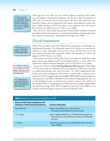
Depending on the onset of VAP, predominant microorganisms include flora of the
upper airway, gram-negative bacilli and methicillin-resistant S. aureus. Table 15-5
outlines the common microbes during the course of VAP (Torres et al., 2004).
clinical pulmonary infection A score of .6 in the modified clinical pulmonary infection score (CPIS) (Table
score: An objective scoring system
to use as an additional aid in the 15-6) has been used as an additional aid in the diagnosis of VAP and in decisions
diagnosis of ventilator-associated on antimicrobial therapy. One study (Fartoukh et al., 2003) emphasized the dif-
pneumonia (VAP) and decision on
antimicrobial therapy. ficulties of the clinical diagnosis of pneumonia in mechanically ventilated patients
suspected of VAP. It suggested that the modified CPIS should be used cautiously in
clinical practice, and further refinements of the clinical scoring approach (e.g., use
A modified CPIS score
of more than 6 at baseline or of other biological markers of infection) are needed to improve the usefulness of
after incorporating the gram the modified CPIS in the management of VAP. Laboratory samples obtained from
stains or culture result is sug-
gestive of pneumonia. bronchoalveolar lavage (BAL) fluid and protected specimen brush (PSB) are two
examples of useful biological markers of infection (Mayhall, 2001).
TABLE 15-5 Common microbes during the Course of VAP
Onset of VAP After Intubation and
Initiation of Mechanical Ventilation Common Microbes
First 48 hours Upper airway flora (e.g., Haemophilus
influenza and Streptococcus pneumonia)
3 to 7 days Gram-negative bacilli (e.g., Pseudomonas aeruginosa,
Escherichia coli, Acinetobacter, Proteus and Klebsiella
species)
.7 days Staphylococcus aureus to include methicillin-resistant
strain (MRSA)
© Cengage Learning 2014
Copyright 2013 Cengage Learning. All Rights Reserved. May not be copied, scanned, or duplicated, in whole or in part. Due to electronic rights, some third party content may be suppressed from the eBook and/or eChapter(s).
Editorial review has deemed that any suppressed content does not materially affect the overall learning experience. Cengage Learning reserves the right to remove additional content at any time if subsequent rights restrictions require it.