
Page 597 - Clinical Application of Mechanical Ventilation
P. 597
Neonatal Mechanical Ventilation 563
intubated immediately after birth. These studies have shown to improve pulmonary
outcome of very-low-birth-weight (VLBW) infants without increasing the incidence
of intraventricular hemorrhage (IVH) and/or periventricular leukomalacia (PVL)
(Durand et al., 2001). Since hyaline membrane disease is the most common condition
in the neonatal ICU, premature infants are most likely to be considered for HFOV
(Vierzig et al., 1994). The indications for HFOV are highly variable and dependent
Infants with congenital on the diagnosis and progression of the patient condition. Infants with congenital di-
diaphragmatic hernia, diffuse aphragmatic hernia (Miguet et al., 1994), diffuse alveolar disease, nonhomogeneous
alveolar disease, nonhomoge-
neous lung disease, air leak, lung disease, air leak, and pulmonary hypoplasia are potential candidates for HFOV.
and pulmonary hypoplasia are
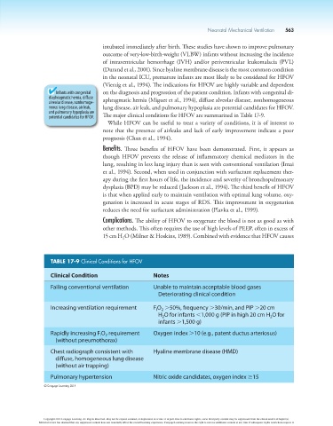
potential candidates for HFOV. The major clinical conditions for HFOV are summarized in Table 17-9.
While HFOV can be useful to treat a variety of conditions, it is of interest to
note that the presence of airleaks and lack of early improvement indicate a poor
prognosis (Chan et al., 1994).
Benefits. Three benefits of HFOV have been demonstrated. First, it appears as
though HFOV prevents the release of inflammatory chemical mediators in the
lung, resulting in less lung injury than is seen with conventional ventilation (Imai
et al., 1994). Second, when used in conjunction with surfactant replacement ther-
apy during the first hours of life, the incidence and severity of bronchopulmonary
dysplasia (BPD) may be reduced (Jackson et al., 1994). The third benefit of HFOV
is that when applied early to maintain ventilation with optimal lung volume, oxy-
genation is increased in acute stages of RDS. This improvement in oxygenation
reduces the need for surfactant administration (Plavka et al., 1999).
Complications. The ability of HFOV to oxygenate the blood is not as good as with
other methods. This often requires the use of high levels of PEEP, often in excess of
15 cm H O (Milner & Hoskins, 1989). Combined with evidence that HFOV causes
2
TABLE 17-9 Clinical Conditions for HFOV
Clinical Condition Notes
Failing conventional ventilation Unable to maintain acceptable blood gases
Deteriorating clinical condition
Increasing ventilation requirement F O .50%, frequency .30/min, and PIP .20 cm
2
I
H O for infants ,1,000 g (PIP in high 20 cm H O for
2
2
infants .1,500 g)
Rapidly increasing F O requirement Oxygen index .10 (e.g., patent ductus arteriosus)
I
2
(without pneumothorax)
Chest radiograph consistent with Hyaline membrane disease (HMD)
diffuse, homogeneous lung disease
(without air trapping)
Pulmonary hypertension Nitric oxide candidates, oxygen index $15
© Cengage Learning 2014
Copyright 2013 Cengage Learning. All Rights Reserved. May not be copied, scanned, or duplicated, in whole or in part. Due to electronic rights, some third party content may be suppressed from the eBook and/or eChapter(s).
Editorial review has deemed that any suppressed content does not materially affect the overall learning experience. Cengage Learning reserves the right to remove additional content at any time if subsequent rights restrictions require it.