
Page 598 - Clinical Application of Mechanical Ventilation
P. 598
564 Chapter 17
hyperinflation of the alveoli, high levels of PEEP may compromise cardiac output
During HFJV and HFOV, and lead to a higher risk of developing barotrauma (Milner & Hoskins, 1989).
cardiopulmonary assessment There are several technical problems encountered in the use of HFOV. One prob-
of the patient is difficult.
Signs of pallor, cyanosis, lem is in the measurement of pressure at the distal end of the endotracheal tube. It
bradycardia, hypotension, and
increased respiratory effort is likely that alveolar pressures are quite different from those measured at the carina.
indicate a worsening of status. An additional problem is a general lack of HFOV devices and training for their use
in level I and level II nurseries.
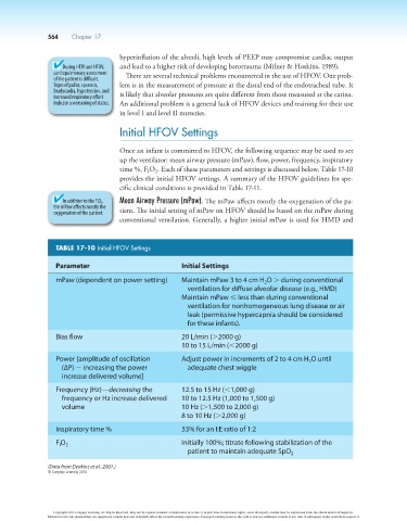
Initial HFOV Settings
Once an infant is committed to HFOV, the following sequence may be used to set
up the ventilator: mean airway pressure (mPaw), flow, power, frequency, inspiratory
time %, F O . Each of these parameters and settings is discussed below. Table 17-10
I
2
provides the initial HFOV settings. A summary of the HFOV guidelines for spe-
cific clinical conditions is provided in Table 17-11.
In addition to the F I O 2 , Mean Airway Pressure (mPaw). The mPaw affects mostly the oxygenation of the pa-
the mPaw affects mostly the
oxygenation of the patient. tient. The initial setting of mPaw on HFOV should be based on the mPaw during
conventional ventilation. Generally, a higher initial mPaw is used for HMD and
TABLE 17-10 Initial HFOV Settings
Parameter Initial Settings
mPaw (dependent on power setting) Maintain mPaw 3 to 4 cm H O . during conventional
2
ventilation for diffuse alveolar disease (e.g., HMD)
Maintain mPaw # less than during conventional
ventilation for nonhomogeneous lung disease or air
leak (permissive hypercapnia should be considered
for these infants).
Bias flow 20 L/min (.2000 g)
10 to 15 L/min (,2000 g)
Power [amplitude of oscillation Adjust power in increments of 2 to 4 cm H O until
2
(∆P) 2 increasing the power adequate chest wiggle
increase delivered volume]
Frequency (Hz)—decreasing the 12.5 to 15 Hz (,1,000 g)
frequency or Hz increase delivered 10 to 12.5 Hz (1,000 to 1,500 g)
volume 10 Hz (.1,500 to 2,000 g)
8 to 10 Hz (.2,000 g)
Inspiratory time % 33% for an I:E ratio of 1:2
F O 2 Initially 100%; titrate following stabilization of the
I
patient to maintain adequate SpO 2
(Data from Deakins et al., 2001.)
© Cengage Learning 2014
Copyright 2013 Cengage Learning. All Rights Reserved. May not be copied, scanned, or duplicated, in whole or in part. Due to electronic rights, some third party content may be suppressed from the eBook and/or eChapter(s).
Editorial review has deemed that any suppressed content does not materially affect the overall learning experience. Cengage Learning reserves the right to remove additional content at any time if subsequent rights restrictions require it.