
Page 78 - Clinical Application of Mechanical Ventilation
P. 78
44 Chapter 2
Ventilatory and Oxygenation Failure
Ventilatory and oxygenation failure has serious and detrimental effects on the
central nervous system (CNS). Such failure may occur in patients on mechani-
cal ventilation because of preexisting clinical conditions, making ventilation
and oxygenation extremely difficult to accomplish in spite of high F O and
2
I
PEEP.
Abnormalities in ventilation and gas exchange can cause hypercapnia (increase in
PaCO ), respiratory acidosis (decrease in pH as a result of the increased PaCO ),
2
2
hypoxemia (decrease in PaO ), secondary polycythemia (increase in red blood cell
2
concentration and thus hemoglobin level), and electrolyte disturbances. These
changes may lead to neurologic impairment.
Indicators of Neurologic Impairment
When neurologic functions are impaired due to ventilatory and oxygenation fail-
Headache, mental status
changes, motor disturbances, ure, the patient may experience headache, mental status changes, motor distur-
and ocular abnormalities bances, and ocular abnormalities (Jozefowicz, 1989).
may be signs of neurologic
impairment. The patient usually describes the headache as “pressure in the head,” having a
higher intensity during night and early morning hours. The headache is the result
of cerebral vasodilation in response to hypoventilation and CO retention during
2
sleep.
Hypoxia, hypercapnia, and acidosis are responsible for the changes in a patient’s
mental status. Early mental disturbances include drowsiness, forgetfulness, and
irritability. In severe or chronic cases of hypoxia and hypercapnia, stupor and coma
may occur.
Hypercapnia may also cause muscle tremor and ocular abnormalities. Muscle
tremor is the result of excessive stimulation of the sympathetic nervous system
and catecholamine release from the adrenal medulla. Ocular abnormalities such as
papilledema, swelling of the area where the optic nerve exits the back of the eye,
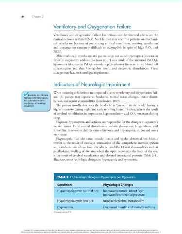
is the result of cerebral vasodilation and elevated intracranial pressure. Table 2-11
illustrates some neurologic changes in hypercapnia and hypoxemia.
TABLE 2-11 Neurologic Changes in Hypercapnia and Hypoxemia
Condition Physiologic Changes
Hypercapnia (with normal pH) Increased cerebral blood flow
Increased intracranial pressure
Hypercapnia (with low pH) Impaired cerebral metabolism
Hypoxemia Decreased mental and motor functions
© Cengage Learning 2014
Copyright 2013 Cengage Learning. All Rights Reserved. May not be copied, scanned, or duplicated, in whole or in part. Due to electronic rights, some third party content may be suppressed from the eBook and/or eChapter(s).
Editorial review has deemed that any suppressed content does not materially affect the overall learning experience. Cengage Learning reserves the right to remove additional content at any time if subsequent rights restrictions require it.