
Page 73 - Clinical Application of Mechanical Ventilation
P. 73
Effects of Positive Pressure Ventilation 39
ABDOMINAL CONSIDERATIONS
Increases in intra-abdominal pressure (IAP) are related to clinical conditions
intra-abdominal pressure
(IAP): Pressure measured by a such as bowel edema or obstruction and ascites. IAP may also be increased in pro-
transducer via a transurethral cedures such as use of pneumatic antishock garments and surgical repair of ab-
bladder catheter.
dominal wall hernias. When these patients are placed on mechanical ventilation,
conditions that are conducive to an increase in IAP should be monitored to avert
potential complications.
Effects of PEEP and Increased
Intra-Abdominal Pressure
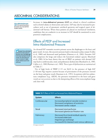
An elevated IAP transmits excessive pressure across the diaphragm to the heart and
Elevated intra-abdominal great vessels. In turn, this excessive pressure leads to decreased cardiac output (Cullen
pressure transmits excessive
pressure across the diaphragm et al., 1989) and decreased renal perfusion (Harman et al., 1982). Excessive IAP
to the heart and great vessels. also compresses the lungs and reduces the functional residual capacity (Burchard
In turn, this excess pressure
leads to decreased cardiac et al., 1985). It has been shown that use of PEEP on patients with elevated IAP
output and decreased renal
perfusion. may lead to cardiovascular, renal, and pulmonary dysfunction (Burchard et al., 1985;
Cullen et al., 1989; Harman et al., 1982). These types of dysfunction are summarized
in Table 2-7.
Use of high levels of PEEP (.15 cm H O) in the presence of high IAP
2
(.20 mm Hg) requires caution because of potentiation of the pressures exerted
on the heart and great vessels (Sussman et al., 1991). In patients with low pulmo-
nary compliance (e.g., ARDS), the pressures transmitted to the heart and great
vessels are not as severe as due to the dampening effects of the noncompliant lungs
and chest wall.
TABLE 2-7 Effects of PEEP and Increased Intra-Abdominal Pressure
System Effects
Cardiovascular Increased peripheral vascular resistance
Decreased compliance of ventricles
Decreased cardiac output
Renal Decreased renal perfusion
Decreased glomerular filtration rate
Pulmonary Decreased functional residual capacity
Increased atelectasis
Impaired gas exchange
Increased V/Q mismatch and venous admixture
© Cengage Learning 2014
Copyright 2013 Cengage Learning. All Rights Reserved. May not be copied, scanned, or duplicated, in whole or in part. Due to electronic rights, some third party content may be suppressed from the eBook and/or eChapter(s).
Editorial review has deemed that any suppressed content does not materially affect the overall learning experience. Cengage Learning reserves the right to remove additional content at any time if subsequent rights restrictions require it.