
Page 105 - Cardiac Nursing
P. 105
1 A
M
9-0
qxd
96.
8:4
P
1 A
pta
ra
g
e 8
p06
0-c
03_
9/2
K34
LWB K34 0-c 03_ p06 9-0 96. qxd 0 9/0 9/2 009 0 0 8:4 1 A M P a a g e 8 1 A pta ra
L L LWB
LWBK340-c03_p069-096.qxd 09/09/2009 08:41 AM Page 81 Aptara
9/0
0
009
C HAPTER 3 / Regulation of Cardiac Output and Blood Pressure 81
200 reflex is caused by pulmonary hyperventilation, which leads to
hypocapnia and activation of pulmonary stretch receptors. The
chemosensor reflex plays only a minimal role in the control of
Δ R-R interval, (msec) 130 eral chemosensor responses may contribute to sympathetic over-
heart rate because the primary and secondary reflexes tend to off-
36
In heart failure, abnormal central and periph-
set one another.
60
164,165
activity and suppression of baroreceptor function.
–10
–80 Respiratory Sinus Arrhythmia
There is a direct relation between heart rate and respiration. Dur-
ing inspiration the heart rate increases, then it decreases during ex-
piration. This respiratory-induced cyclical variation in heart rate is
–150
referred to as a respiratory sinus arrhythmia. There is an ongoing
50 90 130 170 210
debate whether this arrhythmia is due to a central mechanism, a
Carotid distending pressure, (mm/Hg) 166
baroreflex, or a combination of both. The effector arm of this
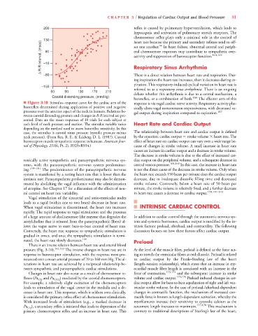
■ Figure 3-10 Stimulus–response curve for the cardiac arm of the response is via vagal cardiac nerve activity. Respiratory activity pha-
baroreflex determined during application of positive and negative sically alters vagal motorneuron responsiveness, with decreased va-
pressures over the anterior aspect of the neck in humans. Relations be- gal output during inspiration compared to expiration. 167
tween carotid distending pressure and changes in R-R interval are pre-
R
R
sented. Data are the mean responses of 10 trials for each subject at
each level of neck pressure and suction. The stimulus variable varies Heart Rate and Cardiac Output
depending on the method used to assess baroreflex sensitivity. In this
case, the stimulus is carotid sinus pressure (systolic pressure minus The relationship between heart rate and cardiac output is defined
neck pressure). (From Rea, R. F., & Eckberg, D. L. (1987). Carotid by the equation: cardiac output stroke volume heart rate. The
baroreceptor-muscle sympathetic response in humans. American Jour- effect of heart rate on cardiac output can vary over a wide range be-
nal of Physiology, 253(6, Pt. 2), R929–R934.) cause of changes in stroke volume. A small increase in heart rate
causes an increase in cardiac output and a decrease in stroke volume.
The decrease in stroke volume is due to the effect of increased car-
tonically active sympathetic and parasympathetic nervous sys- diac output on the peripheral volume, and a subsequent decrease in
tems, with the parasympathetic nervous system predominat- central venous pressure. 168,169 In this case, the increase in heart rate
ing. 159–161 The predominance of the parasympathetic nervous is not the direct cause of the decrease in stroke volume. Only when
system is manifested by a resting heart rate that is lower than the the heart rate exceeds 150 beats per minute does the cardiac output
intrinsic rate. Parasympathetic predominance may also be demon- decrease, due to inadequate diastolic filling time and decreased
strated by abolishing the vagal influence with the administration stroke volume. Conversely, below a heart rate of 50 beats per
of atropine. See Chapter 17 for a discussion of the effects of neu- minute, the stroke volume is relatively fixed, and a further decrease
ral control on heart rate variability. in heart rate causes a decrease in cardiac output. 159,170–172
Vagal stimulation of the sinoatrial and atrioventricular nodes
leads to a rapid (within one to two beats) decrease in heart rate.
When vagal stimulation is discontinued, the heart rate increases INTRINSIC CARDIAC CONTROL
rapidly. The rapid response to vagal stimulation and the presence
of a large amount of cholinesterase (the enzyme that degrades the In addition to cardiac control through the autonomic nervous sys-
acetylcholine that is released from the parasympathetic fibers) al- tem and systemic hormones, cardiac output is modified by the in-
lows the vagus nerve to exert beat-to-beat control of heart rate. trinsic factors: preload, afterload, and contractility. The following
Conversely, the heart rate response to sympathetic stimulation is discussion focuses on how these factors affect cardiac output.
gradual in onset, and once the sympathetic stimulation is termi-
nated, the heart rate slowly decreases. 160 Preload
There is an inverse relation between heart rate and arterial blood
pressure (Fig. 3-10). 162,163 The inverse changes in heart rate are in At the level of the muscle fiber, preload is defined as the force act-
response to baroreceptor stimulation, with the response most pro- ing to stretch the ventricular fibers at end-diastole. Preload is related
nounced over a mean arterial pressure of 70 to 160 mm Hg. The al- to cardiac output by the Frank–Starling law of the heart
terations in heart rate are achieved by a reciprocal relationship be- (length–tension relationship), which states that an increase in my-
tween sympathetic and parasympathetic cardiac stimulations. ocardial muscle fiber length is associated with an increase in the
Changes in heart rate also occur as a result of chemosensor re- force of contraction, 173,174 and the subsequent increase in stroke
P P ) mediated by the carotid chemoreceptors. volume and cardiac output. 175,176 Preload induced changes in car-
P
P
flexes (Pa O 2 and Pa CO 2
For example, a relatively slight excitation of the chemoreceptors diac output allow for beat-to-beat equalization of right and left ven-
leads to stimulation of the vagal center in the medulla and a de- tricular stroke volume. In the case of preload-/afterload-dependent
crease in heart rate. This response, which is seldom seen clinically, changes in contractile function, the mechanism of increased con-
is considered the primary reflex effect of chemosensor stimulation. tractile force is known as length-dependent activation, whereby the
With increased levels of stimulation (e.g., a marked decrease in myofilaments increase their sensitivity to cytosolic calcium as the
), a secondary reflex is initiated that leads to depression of the sarcomere length increases to maximum. 177,178 This mechanism is
Pa P P O 2
primary chemoreceptor reflex and an increase in heart rate. This contrary to traditional descriptions of Starling’s law of the heart,