
Page 159 - Cardiac Nursing
P. 159
LWBK340-c06_p132-152.qxd 09/09/2009 08:25 AM Page 135 Aptara
C HAP TE R 6 / Hematopoiesis, Coagulation, and Bleeding 135
Vascular Spasm
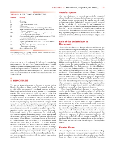
Table 6-2 ■ BLOOD COAGULATION PROTEINS
The sympathetic nervous system is automatically stimulated
Number Name(s) when a blood vessel is injured. Epinephrine and norepinephrine
are released causing contraction of the vascular smooth muscle
I Fibrinogen
II Prothrombin and vasoconstriction. Endothelin I, which is a peptide produced
III Tissue factor (thromboplastin) by the endothelial cell, angiotensin II, and vasoconstrictor
IV Calcium ions prostaglandins are additional agents that contribute to vasocon-
V Proaccelerin, labile factor, or accelerator globulin (AcG) striction. The vasoconstriction of arterioles may be sufficient to
5
VII Serum prothrombin conversion accelerator (SPCA), stable
factor, or proconvertin decrease blood flow and close disrupted capillaries. Larger vessels
VIII Antihemophilic factor (AHF), antihemophilic factor A, may require longer periods of more intense vasoconstriction to
or antihemophilic globulin (AHG) assist with hemostasis, but may ultimately require surgical inter-
IX Christmas factor, plasma thromboplastin component (PTC), vention.
or anthemophilic factor B
X Stuart factor, prower factor, or thrombokinase
XI Plasma thromboplastin antecedent (PTA) or antihemophilic Role of the Endothelium in
factor C
XII Hageman factor, glass factor, contact factor, or Hemostasis
antihemophilic factor D
XIII Fibrin-stabilizing factor (FSF) The endothelial cell was once thought to be inert and have no spe-
cific role in maintaining vascular integrity. Research over the years
has proven this hypothesis to be incorrect. The endothelial cell is
Adapted from Tortora, G., & Grabowski, S. (2003). Principles of Anatomy and Physiol-
ogy. New York: John Wiley & Sons, Inc. a vital component of normal homeostasis. Under normal condi-
tions, the endothelium surface is intact and there is minimal in-
teraction with platelets or the coagulation proteins. The function
of the endothelium is to promote blood flow. The endothelial cell
whose role can be underestimated. To balance the coagulation inhibits blood coagulation by: (1) expressing thrombomodulin, a
process, there are also a number of proteins and systems that will clotting enzyme that binds thrombin; (2) changing the specificity
inhibit coagulation including antithrombin III, proteins C and S, of thrombomodulin from fibrin to protein C, which blocks the
as well as components of the fibrinolytic cascade. The interaction ability to convert fibrinogen to fibrin; (3) using proteoglycans on
of all these proteins in a chemical sequence will produce a clot to their surfaces to bind and potentiate the coagulation inhibitors
repair blood vessels and then dissolve the clot so that normal flow antithrombin III and tissue factor pathway inhibitor; (4) releasing
can be restored. small amounts of plasminogen activator tissue-type plasminogen
activator (tPA); (5) inhibiting platelet aggregation by producing
prostacyclin and nitric oxide, which vasodilates the microcircula-
HEMOSTASIS tion; and (6) inhibiting adherence of peripheral blood cells. 6
These interactions maintain the anticoagulant properties of the
The normal hemostatic system is designed to protect against endothelium by keeping platelets inactive and inhibiting key co-
bleeding from injured blood vessels. Hemostasis is usually ac- agulation proteins such as tissue factor and thrombin.
complished by a sequence of interrelated processes involving Once the endothelial surface is disrupted by various factors, in-
blood vessels and endothelial activity, platelets, and coagulation cluding physical injury or circulating mediators, it will develop
proteins. This complex system is highly regulated to ensure that procoagulant properties. When the endothelium is stimulated by
clotting occurs only at a site of injury and only as long as the in- inflammatory cytokines such as IL -, IL -, or tumor necrosis fac-
tegrity of the vessel is compromised. The process of hemostasis tor , it is referred to as activated endothelium. Once the suben-
consists of several components: (1) blood vessel spasm; (2) for- dothelial connective tissue is exposed and activated, it will lose
mation of a platelet plug; (3) contact between damaged blood thrombomodulin and heparin sulfate and begin to synthesize tis-
vessel, blood platelet, and coagulation proteins; (4) development sue factor (factor III). Factor III interacts with factor VII to start
of a blood clot around the injury; and (5) fibrinolytic removal of the extrinsic pathway. Therefore, protein C is not activated and
5
excess hemostatic material to reestablish vascular integrity. Co- the action of clotting inhibitor systems will be lost. This activation
agulation proteins make up the coagulation cascade. The coagu- of the cascade will further incite the endothelial cell to produce
lation cascade consists of three components: the intrinsic path- more inflammatory mediators (cytokines and chemokines) that
way (vascular trauma), the extrinsic pathway (tissue trauma), and will start the expression of adhesion molecules. Leukocytes will
the common pathway leading to fibrin formation. The clotting adhere to the endothelial cell and become activated by the pro-
processes are balanced by the complex mechanism of fibrinolysis, duction of leukocyte agonists such as platelet activating factor. 6
which breaks down clots and maintains or re-establishes blood Platelets are now attracted to the site and augment the coagulation
flow once the vessel damage has healed. The balance between process.
these two mechanisms and their activators and inhibitors is vital.
An imbalance in one direction leads to excessive bleeding; Platelet Phase
whereas an imbalance in the other direction leads to excessive
clotting. The following sections present the normal sequence of The platelet phase refers to the formation of a soft mass of ag-
coagulation and fibrinolysis, as well as selected coagulation disor- gregated platelets that provides a temporary patch over the in-
ders most commonly associated with the patient experiencing jured vessel. Almost immediately after vascular injury, platelets
cardiovascular disease. begin to adhere to the exposed subendothelial basement