
Page 180 - Cardiac Nursing
P. 180
LWBK340-c07_p153-176.qxd 6/29/09 10:14 PM Page 156 Aptara Inc.
156 PA R T II / Physiologic and Pathologic Responses
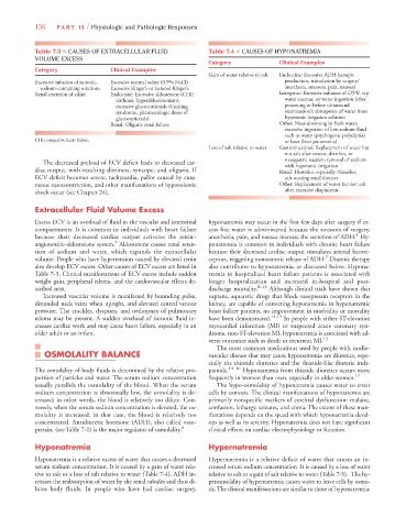
Table 7-3 ■ CAUSES OF EXTRACELLULAR FLUID Table 7-4 ■ CAUSES OF HYPONATREMIA
VOLUME EXCESS
Category Clinical Examples
Category Clinical Examples
Gain of water relative to salt Endocrine: Excessive ADH (ectopic
Excessive infusion of isotonic, Excessive normal saline (0.9% NaCl) production; stimulation by surgery/
sodium-containing solutions Excessive Ringer’s or lactated Ringer’s anesthesia, stressors, pain, nausea)
Renal retention of saline Endocrine: Excessive aldosterone (CHF, Iatrogenic: Excessive infusion of D5W, tap
cirrhosis, hyperaldosteronism); water enemas, or water ingestion (after
excessive glucocorticoids (Cushing poisoning or before ultrasound
syndrome, pharmacologic doses of examination); absorption of water from
glucocorticoids) hypotonic irrigation solution
Renal: Oliguric renal failure Other: Near-drowning in fresh water;
excessive ingestion of low-sodium fluid
such as water (psychogenic polydipsia)
CHF, congestive heart failure. or beer (beer potomania)
Loss of salt relative to water Gastrointestinal: Replacement of water but
not salt after emesis, diarrhea, or
nasogastric suction; removal of sodium
The decreased preload of ECV deficit leads to decreased car- with hypotonic irrigation
diac output, with resulting dizziness, syncope, and oliguria. If Renal: Diuretics, especially thiazides;
ECV deficit becomes severe, tachycardia, pallor caused by cuta- salt-wasting renal diseases
neous vasoconstriction, and other manifestations of hypovolemic Other: Replacement of water but not salt
shock occur (see Chapter 24). after excessive diaphoresis
Extracellular Fluid Volume Excess
Excess ECV is an overload of fluid in the vascular and interstitial hyponatremia may occur in the first few days after surgery if ex-
compartments. It is common in individuals with heart failure cess free water is administered because the stressors of surgery,
1
because their decreased cardiac output activates the renin– anesthesia, pain, and nausea increase the secretion of ADH. Hy-
5
angiotensin–aldosterone system. Aldosterone causes renal reten- ponatremia is common in individuals with chronic heart failure
tion of sodium and water, which expands the extracellular because their decreased cardiac output stimulates arterial barore-
7
volume. People who have hypertension caused by elevated renin ceptors, triggering nonosmotic release of ADH. Diuretic therapy
also develop ECV excess. Other causes of ECV excess are listed in also contributes to hyponatremia, as discussed below. Hypona-
Table 7-3. Clinical manifestations of ECV excess include sudden tremia in hospitalized heart failure patients is associated with
weight gain, peripheral edema, and the cardiovascular effects de- longer hospitalization and increased in-hospital and post-
scribed next. discharge mortality. 8–10 Although clinical trials have shown that
Increased vascular volume is manifested by bounding pulse, vaptans, aquaretic drugs that block vasopressin receptors in the
distended neck veins when upright, and elevated central venous kidney, are capable of correcting hyponatremia in hyponatremic
pressure. The crackles, dyspnea, and orthopnea of pulmonary heart failure patients, no improvement in morbidity or mortality
edema may be present. A sudden overload of isotonic fluid in- have been demonstrated. 11,12 In people with either ST-elevation
creases cardiac work and may cause heart failure, especially in an myocardial infarction (MI) or suspected acute coronary syn-
older adult or an infant. drome, non-ST-elevation MI, hyponatremia is associated with ad-
verse outcomes such as death or recurrent MI. 13
The most common medications used by people with cardio-
OSMOLALITY BALANCE vascular disease that may cause hyponatremia are diuretics, espe-
cially the thiazide diuretics and the thiazide-like diuretic inda-
The osmolality of body fluids is determined by the relative pro- pamide. 14–16 Hyponatremia from thiazide diuretics occurs more
portion of particles and water. The serum sodium concentration frequently in women than men, especially in older women. 17
usually parallels the osmolality of the blood. When the serum The hypo-osmolality of hyponatremia causes water to enter
sodium concentration is abnormally low, the osmolality is de- cells by osmosis. The clinical manifestations of hyponatremia are
creased; in other words, the blood is relatively too dilute. Con- primarily nonspecific markers of cerebral dysfunction: malaise,
versely, when the serum sodium concentration is elevated, the os- confusion, lethargy, seizures, and coma. The extent of these man-
molality is increased; in that case, the blood is relatively too ifestations depends on the speed with which hyponatremia devel-
concentrated. Antidiuretic hormone (ADH), also called vaso- ops as well as its severity. Hyponatremia does not have significant
pressin, (see Table 7-1) is the major regulator of osmolality. 6 clinical effects on cardiac electrophysiology or function.
Hyponatremia Hypernatremia
Hyponatremia is a relative excess of water that causes a decreased Hypernatremia is a relative deficit of water that causes an in-
serum sodium concentration. It is caused by a gain of water rela- creased serum sodium concentration. It is caused by a loss of water
tive to salt or a loss of salt relative to water (Table 7-4). ADH in- relative to salt or a gain of salt relative to water (Table 7-5). The hy-
creases the reabsorption of water by the renal tubules and thus di- perosmolality of hypernatremia causes water to leave cells by osmo-
lutes body fluids. In people who have had cardiac surgery, sis. The clinical manifestations are similar to those of hyponatremia: