
Page 179 - Cardiac Nursing
P. 179
LWBK340-c07_p153-176.qxd 6/29/09 10:14 PM Page 155 Aptara Inc.
C HAP TE R 7 / Fluid and Electrolyte and Acid–Base Balance and Imbalance 155
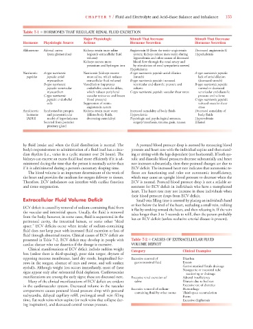
Table 7-1 ■ HORMONES THAT REGULATE RENAL FLUID EXCRETION
Major Physiologic Stimuli That Increase Stimuli That Decrease
Hormone Physiologic Source Actions Hormone Secretion Hormone Secretion
Aldosterone Adrenal cortex Kidneys retain more saline Angiotensin II (from the renin–angiotensin Decreased angiotensin II
(zona glomerulosa) (expands extracellular fluid system; kidneys release more renin during Hyperkalemia
volume) hypovolemia and other causes of decreased
Kidneys excrete more blood flow through the renal artery and
potassium and hydrogen ions by stimulation of renal sympathetic nerves)
Hypokalemia
Natriuretic A-type natriuretic Natriuresis (kidneys excrete A-type natriuretic peptide: atrial dilation A-type natriuretic peptide:
peptides peptide: atrial more saline, which reduces (stretch) lack of atrial dilation
myocardium extracellular fluid volume) B-type natriuretic peptide: increased (decreased stretch)
B-type natriuretic Vasodilation (suppresses ventricular end-diastolic pressure and B-type natriuretic peptide:
peptide: ventricular endothelin; arterioles dilate, volume normal or decreased
myocardium which reduces peripheral C-type natriuretic peptide: vascular shear stress ventricular end-diastolic
C-type natriuretic vascular resistance and lowers pressure and volume
peptide: endothelial blood pressure) C-type natriuretic peptide:
cells Suppression of renin– reduced vascular shear
angiotensin system stress
Antidiuretic Synthesized in preoptic Kidneys retain more water Increased osmolality of body fluids Decreased osmolality of
hormone and paraventricular (dilutes body fluids, Hypovolemia body fluids
(ADH) nuclei of hypothalamus decreasing osmolality) Physiologic and psychological stressors, Hypervolemia
Secreted from posterior surgery/anesthesia, trauma, pain, nausea Ethanol
pituitary gland
by fluid intake and when the fluid distribution is normal. The A postural blood pressure drop is assessed by measuring blood
body’s responsiveness to administration of a fluid load has a circa- pressure and heart rate with the individual supine and then stand-
dian rhythm (i.e., varies in a cyclic manner over 24 hours). The ing or sitting with the legs dependent (not horizontal). If both sys-
kidneys can excrete an excess fluid load more efficiently if it is ad- tolic and diastolic blood pressures decrease substantially and heart
ministered during the time that the person is normally active than rate increases substantially, then these postural changes are due to
if it is administered during a person’s customary sleeping time. ECV deficit. The increased heart rate indicates that autonomic re-
The blood volume is an important determinant of the work of flexes are functioning and rules out autonomic insufficiency,
the heart and provides the medium for oxygen delivery to tissues. which may cause an upright blood pressure to decrease when the
Therefore, ECV imbalances can interfere with cardiac function ECV is normal. Postural blood pressure drop is not a reliable as-
and tissue oxygenation. sessment for ECV deficit in individuals who have a transplanted
heart. The heart rate may not increase in these individuals when
their blood pressure drops from ECV deficit.
Extracellular Fluid Volume Deficit Small vein filling time is assessed by placing an individual’s hand
or foot below the level of the heart, occluding a small vein, milking
ECV deficit is caused by removal of sodium-containing fluid from it flat by stroking toward the heart, and then releasing it. If the vein
the vascular and interstitial spaces. Usually, the fluid is removed takes longer than 3 to 5 seconds to refill, then the person probably
from the body; however, in some cases, fluid is sequestered in the has an ECV deficit (unless occlusive arterial disease is present).
peritoneal cavity, the intestinal lumen, or some other “third
space.” ECV deficits occur when intake of sodium-containing
fluid does not keep pace with increased fluid excretion or loss of
fluid through abnormal routes. Clinical causes of ECV deficit are
presented in Table 7-2. ECV deficit may develop in people with Table 7-2 ■ CAUSES OF EXTRACELLULAR FLUID
cardiac disease who use diuretics if the dosage is excessive. VOLUME DEFICIT
Clinical manifestations of ECV deficit include sudden weight Category Clinical Examples
loss (unless there is third-spacing), poor skin turgor, dryness of
opposing mucous membranes, hard dry stools, longitudinal fur- Excessive removal of Diarrhea
rows in the tongue, absence of tears and sweat, and soft sunken gastrointestinal fluid Emesis
eyeballs. Although weight loss occurs immediately, most of these Gastrointestinal fistula drainage
Nasogastric or intestinal tube
signs appear only after substantial fluid depletion. Cardiovascular suctioning or drainage
manifestations are among the early signs; these are discussed next. Excessive renal excretion of Adrenal insufficiency
Many of the clinical manifestations of ECV deficit are evident saline Diuresis due to bed rest
in the cardiovascular system. Decreased volume in the vascular Excessive use of diuretics
compartment causes postural blood pressure drop with postural Excessive removal of sodium- Hemorrhage
containing fluid by other routes
Third-space accumulation
tachycardia, delayed capillary refill, prolonged small vein filling Burns
time, flat neck veins when supine (or neck veins that collapse dur- Excessive diaphoresis
ing inspiration), and decreased central venous pressure.