
Page 183 - Cardiac Nursing
P. 183
LWBK340-c07_p153-176.qxd 6/29/09 10:14 PM Page 159 Aptara Inc.
C HAP TE R 7 / Fluid and Electrolyte and Acid–Base Balance and Imbalance 159
4.0 mEq/L are necessary in individuals who have cardiac arrhyth-
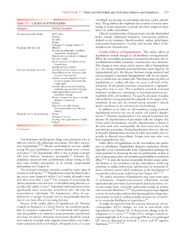
Table 7-7 ■ CAUSES OF HYPOKALEMIA mias. The guidelines also emphasize the necessity of routine mon-
itoring of serum potassium in people who have congestive heart
Category Clinical Examples
failure or cardiac arrhythmias.
Decreased potassium intake NPO orders Clinical manifestations of hypokalemia include diminished
Anorexia bowel sounds, abdominal distention, constipation, polyuria,
Fad diets skeletal muscle weakness, flaccid paralysis, cardiac arrhythmias,
Fasting
Prolonged IV therapy without K
and postural hypotension. Cardiac and vascular effects of hy-
Potassium shift into cells Alkalosis pokalemia are discussed next.
Excessive 2 -adrenergic stimulation
(epinephrine, -agonists) Cardiac Effects of Hypokalemia. The cardiac effects of
Hypothermia (accidental or induced) hypokalemia include changes in cell membrane resting potential.
Excessive insulin When the extracellular potassium concentration decreases, the ex-
Rapid correction of acidosis during tracellular/intracellular potassium concentration ratio decreases.
hemodialysis This change in ratio causes cardiac muscle cells to hyperpolarize
Familial periodic paralysis
Increased potassium excretion Diarrhea (includes laxative overuse) (i.e., the resting membrane potential becomes more negative). In
Hyperaldosteronism (increases renal hyperpolarized cells, the distance between resting potential and
excretion of potassium) action potential is increased; hyperpolarized cells are less respon-
Chronic excessive ingestion of black sive to stimuli than are normal cells. The hyperpolarizing effect of
licorice (contains aldosterone-like
compounds) hypokalemia on cardiac cells does not occur at all levels of hy-
Excessive glucocorticoids (Cushing pokalemia. At low plasma potassium concentrations, a hypopolar-
syndrome; glucocorticoid therapy) izing effect may be seen. This is probably caused by decreased
Hypomagnesemia (causes renal potassium conductance (analogous to decreased potassium per-
potassium wasting) meability) of the cell membrane. The specific alteration of cardiac
Diuretic therapy with loop or thiazide
diuretics or mannitol cell membrane resting potential thus depends on the degree of hy-
Polyuria pokalemia. In any case, the normal resting potential is altered,
High-dose penicillin therapy which contributes to the development of arrhythmias.
(nonreabsorbable anion effect in In addition to its effect on cell membrane resting potential,
kidney)
Potassium loss by abnormal route Emesis hypokalemia increases the rate of cardiac cell diastolic depolar-
29
Nasogastric suction ization. Diastolic depolarization is the normal mechanism that
Drainage from gastrointestinal fistula initiates the depolarization of pacemaker cells (see Chapter 16).
Dialysis Under usual circumstances, diastolic depolarization is fastest in
the sinus node cells; consequently, the sinus node serves as the
IV, intravenous. predominant pacemaker. During hypokalemia, however, the rate
of diastolic depolarization increases in other myocardial cells, es-
pecially in diseased myocardium. Ectopic beats may arise, even
Catecholamines and -agonist drugs cause potassium ions to from hyperpolarized cells.
shift into cells by a 2 -adrenergic mechanism. This effect can pro- Other effects of hypokalemia on the myocardium also predis-
duce hypokalemia. 22,23 Plasma catecholamines increase rapidly pose to arrhythmias. Hypokalemia decreases conduction velocity,
during MI and hypokalemia is common during acute coronary especially in the atrioventricular node. Hypokalemia prolongs the
syndromes. 24 This hypokalemic effect is not as strong in people action potential by decreasing the rate of repolarization, at least in
who have diabetic autonomic neuropathy. 24 Transient hy- part by decreasing cardiac cell membrane permeability to potassium
pokalemia associated with catecholamine release during an MI efflux. 30,31 It alters the normal relationship between action poten-
may cause further impairment of an already compromised tial duration in the epicardium and the endocardium, which may
myocardium (see Chapter 5). contribute to cardiac arrhythmias, and decreases the ventricular ef-
The increased potassium excretion caused by many types of fective refractory period, which predisposes to the development of
diuretics is well known. 21,25 Hypokalemia caused by diuretic ther- extrasystoles and reentrant arrhythmias (see Chapter 16). 31–33
apy occurs most frequently within 2 to 8 weeks, although it may The cardiac alterations of hypokalemia may cause many types
arise after more than 1 year. 26 The necessity of monitoring the of arrhythmias. Hypokalemia-induced arrhythmias include
plasma potassium concentration in individuals using diuretics, es- supraventricular premature depolarizations and tachycardias, ven-
27
pecially older adults, is clear. Individuals with hypokalemia have tricular ectopic beats, ventricular tachycardia, torsade de pointes,
significantly more ventricular arrhythmias after MI than do and ventricular fibrillation. 34–39 Hypokalemia potentiates digitalis
normokalemic individuals. The hypokalemic effect of cate- toxicity. Animal studies indicate that downregulation of gap junc-
cholamines is stronger in people who are using thiazide diuretics tion proteins in diabetic cardiomyopathy increases the vulnerabil-
than it is in those who are not using diuretics. ity to ventricular fibrillation in hypokalemia. 40
Because of the cardiac effects of hypokalemia, the National As might be expected from the previous discussion, electro-
Council on Potassium in Clinical Practice has established guide- cardiographic (ECG) changes are seen in individuals with
lines for potassium replacement. 28 For individuals with hyperten- hypokalemia (see Chapter 16). A characteristic change is the de-
sion, the guideline is to maintain a serum potassium concentration velopment of U waves. 41,42 Other ECG changes include in-
of at least 4.0 mEq/L. Potassium replacement should be consid- creased amplitude of P waves, prolonged PR interval, prolonged
ered routinely in people with congestive heart failure, even with a QT interval, flattened or inverted T waves, and ST segment
serum potassium level of 4.0 mEq/L. Potassium levels of at least depression. 23,32,35,36,42,43