
Page 427 - Cardiac Nursing
P. 427
P
:57
/09
M
a
a
P
19.
0-4
p40
19.
/30
6
qxd
K34
0-c
18_
L L LWB
LWBK340-c18_18_p400-419.qxd 6/30/09 7:57 PM Page 403 Aptara Inc.
K34
LWB K34 0-c 18_ p40 0-4 19. qxd 6 /30 /09 7 7 :57 P M P a g g e e 40 3 A pta ra Inc . .
pta
3 A
40
Inc
ra
pta
C HAPTER 1 8 / Cardiac Electrophysiology Procedures 403
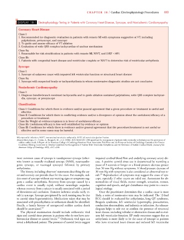
DISPLAY 18-1 Electrophysiology Testing in Patients with Coronary Heart Disease, Syncope, and Nonischemic Cardiomyopathy
Coronary Heart Disease
Class I:
1. Recommended for diagnostic evaluation in patients with remote MI with symptoms suggestive of VT, including
palpitations, presyncope, and syncope
2. To guide and assess efficacy of VT ablation
3. Evaluation of wide QRS complex tachycardias of unclear mechanism
Class IIa:
1. Reasonable for risk stratification in patients with remote MI, NSVT, and LVEF 40%
Class IIb:
1. Patients with congenital heart disease and ventricular couplets or NSVT to determine risk of ventricular arrhythmia
Syncope
Class I:
1. Syncope of unknown cause with impaired left ventricular function or structural heart disease
Class IIa:
1. Syncope with suspected brady or tachyarrhythmia in whom noninvasive diagnostic studies are not conclusive
Nonischemic Cardiomyopathy
Class I:
1. Diagnose bundle-branch reentrant tachycardia and to guide ablation sustained palpitations, wide QRS complex tachycar-
dia, syncope, or presyncope
Classification
Class I: Conditions for which there is evidence and/or general agreement that a given procedure or treatment is useful and
effective.
Class II: Conditions for which there is conflicting evidence and/or a divergence of opinion about the usefulness/efficacy of a
procedure or treatment.
Class IIa: Weight of evidence/opinion is in favor of usefulness/efficacy
Class IIb: Usefulness/efficacy is less well established by evidence or opinion
Class III: Conditions for which there is evidence and/or general agreement that the procedure/treatment is not useful or
effective and in some cases may be harmful.
MI, myocardial infarction; NSVT, nonsustained ventricular tachycardia; LVEF, left ventricular ejection fraction.
Adapted from Zipes, D., Camm, A., Borggrefe, M., et al. (2006). ACC/AHA/ESC 2006 guidelines for management of patients with ventricular arrhythmias and the prevention of
sudden cardiac death: A Report of the American College of Cardiology/American Heart Association Task Force and the European Society of Cardiology Committee for Practice
Guidelines (Writing Committee to Develop Guidelines for Management of Patients With Ventricular Arrhythmias and the Prevention of Sudden Cardiac Death). Journal of the
American College of Cardiology, 48(5), e247–e346.
most common cause of syncope is vasodepressor syncope (other- impaired cerebral blood flow and underlying coronary artery dis-
wise known as neurally mediated syncope (NMS), neurocardio- ease. A positive carotid sinus test is documented by recording a
genic syncope, or vasovagal syncope) followed by primary pause of 3 seconds or longer or a blood pressure decrease greater
arrhythmias. than 50 mm Hg without symptoms. A blood pressure decrease of
The history, including observers’ statements describing the on- 30 mm Hg with symptoms is also considered an abnormal test re-
set and recovery can provide clues for the cause. For example, sud- sult. 25 Reproduction of symptoms may suggest the cause of syn-
den onset of syncope without any warning signs or symptoms sug- cope, especially if other causes are ruled out. Assessment for ab-
gests a cardiac arrhythmia. Recovery from syncope caused by a normalities of visual fields, motor strength, sensation, tremor,
cardiac event is usually rapid, without neurologic sequelae, cognition and speech, and gait disturbance may point to a neuro-
whereas recovery from a seizure is usually associated with a period logical etiology.
of drowsiness and confusion. Transient ischemic attacks rarely re- Once the practitioner determines that a cardiac cause is most
sult in syncope. Syncope precipitated by neck turning may be due likely, a series of noninvasive tests may be indicated. The 12-lead
to carotid sinus hypersensitivity. Medications taken that may be ECG should be evaluated for arrhythmias, long QT syndrome,
associated with proarrhythmia or orthostasis should be identified. Brugada syndrome, left ventricular hypertrophy, preexcitation,
Finally, a family history of unexpected sudden cardiac death conduction abnormalities, and ischemia or infarction. An echocar-
should be ascertained. 23 diogram helps to rule out or confirm the presence of structural
The physical examination should include orthostatic vital heart disease, including valvular or obstructive disease and to eval-
signs and carotid sinus pressure in patients who do not have cere- uate left ventricular function. EP study outcomes suggest that an
brovascular disease or carotid bruits. 24 Orthostatic vital signs can arrhythmia is more likely to be the cause of syncope in patients
reveal a dehydrated patient. The presence of carotid bruits suggest who have structural heart disease and reduced left ventricular