
Page 480 - Cardiac Nursing
P. 480
LWBK340-c20_p439-459.qxd 6/29/09 11:29 PM Page 456 Aptara Inc.
456 PA R T III / Assessment of Heart Disease
tingling may indicate reduced perfusion and must be carefully Temperature
evaluated. A diminished or absent pulse is a sign of serious arterial Early increases in temperature may occur because of the fluid loss
occlusion, which often constitutes a surgical emergency. The first that occurs with catheterization. More persistent elevations may
step, if any of these signs occur, is to check the compression de- indicate infection or pyrogenic reactions.
vice (if used) and release pressure. If symptoms do not resolve, the
physician should be notified immediately and steps should be Urinary Output
taken to preserve the limb. Because angiographic contrast medium acts as an osmotic di-
Manual pressure or pressure with a compression device such as uretic, patients have an increase in urine output for a short time
a FemoStop is used for hemostasis at the time of sheath removal after catheterization. IV fluids are often continued for a variable
and when bleeding continues or recurs after initial hemostasis. time after the procedure, and oral fluids should be encouraged un-
When pressure is applied at an arterial site, the pulse distal to the less the patient has been ordered nothing by mouth for some rea-
site may be safely occluded for 2 to 5 minutes, and then pressure son.
is released until the pulse returns. Distal pulses should remain
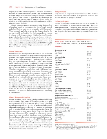
palpable during the remainder of pressure application, which con-
tinues for 15 to 20 minutes. If oozing from the sheath insertion
tract continues after initial hemostasis, infiltration of the tract Table 20-2 ■ NORMAL ADULT VALUES FOR DATA
with a solution of lidocaine and epinephrine (1:100,000 strength) COLLECTED DURING CARDIAC CATHETERIZATION
followed by 2 to 5 minutes of light manual pressure is usually ef-
fective to control bleeding. Pressures mm Hg
Systemic arterial
Blood Pressure Peak-systolic 100–140
Evaluation of the blood pressure after cardiac catheterization End-diastolic 60–90
should include comparison of preprocedure and postprocedure Mean 70–105
pressures, checking for orthostatic hypotension once the bed rest Left ventricular
period is over, and monitoring for paradoxical pulse. Mild sys- Peak-systolic 100–140
End-diastolic 3–12
tolic hypotension frequently occurs after cardiac catheterization
Left atrial
and is usually not of concern. Angiographic contrast medium
Left atrial mean (or PAWP) 1–10
acts as an osmotic diuretic, and patients frequently return with a wave 3–15
signs of volume depletion, including orthostatic hypotension. v wave 3–12
Therefore, patients are kept on bed rest until fluid balance is re- Pulmonary artery
stored with oral liquids or by IV replacement. Hypotension may Peak-systolic 15–30
also be a response to the drugs given during the procedure. If the End-diastolic 3–12
Systolic Mean 9–16
blood pressure is consistently low, other causes need to be inves-
tigated, such as possible blood loss or arrhythmias. Patient assess- Right ventricular
Peak-systolic 15–30
ment needs to be performed and the physician notified. Para-
End-diastolic 0–8
doxical pulse suggests pericardial tamponade, which is very rare
Right atrial
but may occur as a result of perforation of a coronary artery or Mean 8–10
the myocardium. In patients with known perforation, this sign a wave 2–10
should be specifically assessed with each blood pressure measure- v wave 2–10
ment, and, if it occurs, the physician should be notified. Hyper- Left Ventricular Volumes
2
tension can also occur and may contribute to access site bleeding End-systolic volume (mL/m ) 20–30
2
End-diastolic volume (mL/m ) 70–79
if not controlled.
Ejection fraction .58–.72
Resistance (dynes/s/cm 5 5 5 )
Heart Rate and Rhythm Total systemic resistance 900–1,440
Patients who have had an interventional procedure should be on Pulmonary arteriolar (vascular) resistance 37–97
a cardiac monitor for rhythm and ST-segment monitoring. A mild Flow
sinus tachycardia (100 to 120 beats per minute) is not unusual af- CO (L/min) 2 4.0–8.0
Cardiac index (L/min/m )
2.5–4.0
ter catheterization and may be a sign of anxiety, an indication of Stroke index (mL/beat/m ) 35–70
2
saline and water loss due to diuresis, or a reaction to medication Stroke volume (mL/beat) 60–130
2
such as atropine. Fluids, time, and reassurance often bring the Oxygen consumption (mL/min/m ) 125
heart rate down to more normal levels. Heart rates above 120 Oxygen Saturation (%)
beats per minute should be evaluated for other causes such as Right atrium 60–75
hemorrhage, more severe fluid imbalance, fever, or arrhythmias. Right ventricle 60–75
60–75
Pulmonary artery
Bradycardia may indicate vasovagal responses, arrhythmias, or in- Left atrium 95–99
farction and should be assessed by 12-lead ECG and correlated Left ventricle 95–99
with other clinical signs, such as pain and blood pressure. Vasova- Aorta 95–99
gal reactions are fairly common and can occur immediately or
hours after sheath removal. Cardiac monitoring for ST-segment PAWP, pulmonary artery wedge pressure.
From Kucher, N., & Goldhaber, S. Z. (2006). Pulmonary angiography. In Baim, D. S.
displacement is useful to detect acute reocclusion of the artery or (Ed.), Grossman’s cardiac catheterization, angiography, and intervention (7th ed., p.
MI after an interventional procedure. 39,40 236). Philadelphia: Lippincott Williams & Wilkins.