
Page 476 - Cardiac Nursing
P. 476
M
Pa
M
9 P
9 P
g
g
g
Pa
Pa
1:2
/09
/09
/29
6
/29
1
1:2
1
/09
1
a
a
a
ara
ara
c.
c.
In
a
In
t
52
A
52
e 4
e 4
p
t
p
A
p
6
LWB K34 0-c 20_ pp439-459.qxd 6/29/09 11:29 PM Page 452 Aptara Inc.
LWB
p
43
p
20_
20_
0-c
LWBK340-c20_
K34
43
q
q
q
xd
xd
9-4
9-4
59.
59.
59.
452 P A R T III / Assessment of Heart Disease
A
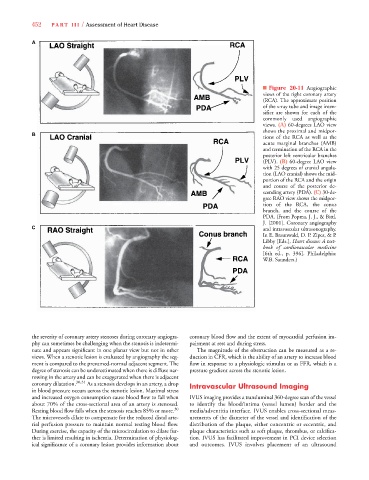
■ Figure 20-11 Angiographic
views of the right coronary artery
(RCA). The approximate position
of the x-ray tube and image inten-
sifier are shown for each of the
commonly used angiographic
views. (A) 60-degrees LAO view
shows the proximal and midpor-
B
tions of the RCA as well as the
acute marginal branches (AMB)
and termination of the RCA in the
posterior left ventricular branches
(PLV). (B) 60-degree LAO view
with 25 degrees of cranial angula-
tion (LAO cranial) shows the mid-
portion of the RCA and the origin
and course of the posterior de-
scending artery (PDA). (C) 30-de-
gree RAO view shows the midpor-
tion of the RCA, the conus
branch, and the course of the
PDA. (From Popma, J. J., & Bittl,
J. [2001]. Coronary angiography
C and intravascular ultrasonography.
In E. Braunwald, D. P. Zipes, & P.
Libby [Eds.], Heart disease: A text-
book of cardiovascular medicine
[6th ed., p. 396]. Philadelphia:
W.B. Saunders.)
the severity of coronary artery stenoses during coronary angiogra- coronary blood flow and the extent of myocardial perfusion im-
phy can sometimes be challenging when the stenosis is indetermi- pairment at rest and during stress.
nate and appears significant in one planar view but not in other The magnitude of the obstruction can be measured as a re-
views. When a stenotic lesion is evaluated by angiography the seg- duction in CFR, which is the ability of an artery to increase blood
ment is compared to the presumed-normal adjacent segment. The flow in response to a physiologic stimulus or as FFR, which is a
degree of stenosis can be underestimated when there is diffuse nar- pressure gradient across the stenotic lesion.
rowing in the artery and can be exaggerated when there is adjacent
coronary dilatation. 30,31 As a stenosis develops in an artery, a drop Intravascular Ultrasound Imaging
in blood pressure occurs across the stenotic lesion. Maximal stress
and increased oxygen consumption cause blood flow to fall when IVUS imaging provides a transluminal 360-degree scan of the vessel
about 70% of the cross-sectional area of an artery is stenosed. to identify the blood/intima (vessel lumen) border and the
Resting blood flow falls when the stenosis reaches 85% or more. 30 media/adventitia interface. IVUS enables cross-sectional meas-
The microvessels dilate to compensate for the reduced distal arte- urements of the diameter of the vessel and identification of the
rial perfusion pressure to maintain normal resting blood flow. distribution of the plaque, either concentric or eccentric, and
During exercise, the capacity of the microcirculation to dilate fur- plaque characteristics such as soft plaque, thrombus, or calcifica-
ther is limited resulting in ischemia. Determination of physiolog- tion. IVUS has facilitated improvement in PCI device selection
ical significance of a coronary lesion provides information about and outcomes. IVUS involves placement of an ultrasound