
Page 475 - Cardiac Nursing
P. 475
1:2
1:2
9 P
/09
1
1
Pa
Pa
g
9 P
M
M
q
xd
xd
59.
q
q
/29
/29
/09
6
6
6
ara
a
a
t
ara
ara
In
c.
c.
a
a
In
e 4
51
51
g
g
e 4
p
p
t
A
A
p
LWBK340-c20_
LWB
LWB K34 0-c 20_ p pp439-459.qxd 6/29/09 11:29 PM Page 451 Aptara Inc.
20_
0-c
K34
9-4
9-4
59.
43
p
43
A A
B B
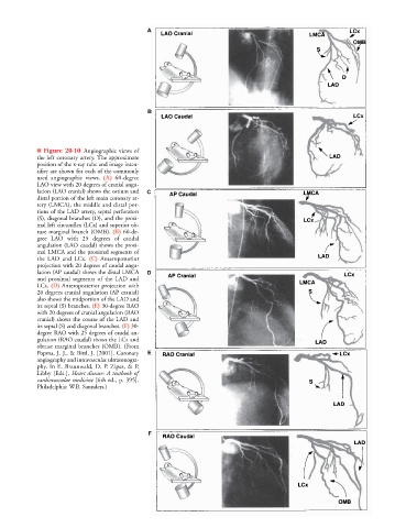
■ Figure 20-10 Angiographic views of
the left coronary artery. The approximate
position of the x-ray tube and image inten-
sifier are shown for each of the commonly
used angiographic views. (A) 60-degree
LAO view with 20 degrees of cranial angu-
lation (LAO cranial) shows the ostium and C
distal portion of the left main coronary ar-
tery (LMCA), the middle and distal por-
tions of the LAD artery, septal perforators
(S), diagonal branches (D), and the proxi-
mal left circumflex (LCx) and superior ob-
tuse marginal branch (OMB). (B) 60-de-
gree LAO with 25 degrees of caudal
angulation (LAO caudal) shows the proxi-
mal LMCA and the proximal segments of
the LAD and LCx. (C) Anteroposterior
projection with 20 degrees of caudal angu-
lation (AP caudal) shows the distal LMCA D
and proximal segments of the LAD and
LCx. (D) Anteroposterior projection with
20 degrees cranial angulation (AP cranial)
also shows the midportion of the LAD and
its septal (S) branches. (E) 30-degree RAO
with 20 degrees of cranial angulation (RAO
cranial) shows the course of the LAD and
its septal (S) and diagonal branches. (F) 30-
degree RAO with 25 degrees of caudal an-
gulation (RAO caudal) shows the LCx and
obtuse marginal branches (OMB). (From
Popma, J. J., & Bittl, J. [2001]. Coronary E E
angiography and intravascular ultrasonogra-
phy. In E. Braunwald, D. P. Zipes, & P.
Libby [Eds.], Heart disease: A textbook of
cardiovascular medicine [6th ed., p. 395].
Philadelphia: W.B. Saunders.)
F F