
Page 635 - Cardiac Nursing
P. 635
LWBK340-c25_p595-622.qxd 06/30/2009 17:45 Page 611 Aptara
C HAPTER 2 5 / Cardiac Surgery 611
potential recipient has cytotoxic antibodies that will react to
that specific donor heart. Table 25-4 ■ INTERNATIONAL SOCIETY OF HEART AND
Acute rejection is the most frequently occurring form of rejec- LUNG TRANSPLANTATION STANDARD GRADING OF
tion and is a major cause of death within the first year after trans- CARDIAC REJECTION
plantation. 54 Preoperative immunosuppressive therapy is begun Revised
for prevention of acute cardiac rejection. Routine monitoring for Grade (2004) Old Grade Nomenclature
acute rejection is centered around endomyocardial biopsy. With
cyclosporine/tacrolimus therapy, there are few clinically evident 0R 0 No rejection
signs and symptoms of acute rejection. The objective is to detect 1R 1 A, Focal, mild
acute rejection in its early stages at a time when the process can be 2 B, Diffuse, mild
One aggressive infiltrate, focal moderate
reversed, thus preventing serious, permanent damage to the new 2R 3 A, Multifocal aggressive, moderate
heart. Therefore, biopsy remains the gold standard for monitoring 3R B, Diffuse inflammatory process
and early detection of acute rejection. Because acute rejection is 4 Diffuse, aggressive, with necrosis, severe
expected to occur during the first 3 months after surgery, biopsy acute rejection
is performed within the first 14 days after transplantation, and
then up to once per week during this crucial time interval. Any
time that rejection is detected, biopsies are performed frequently
to monitor the progress of antirejection treatment. By 1 month,
the biopsy schedule is tapered to every other week, then once per myocyte and vascular necrosis with hemorrhage and a mixed in-
month after the third month. Patients are then monitored indefi- filtrate of immunoblasts and neutrophils ISHLT grade 3R (old
nitely by biopsy every 4 months to annually, depending on the scales 3B and 4). 71 Resolving rejection is evidenced by active fi-
transplant center. Many centers stop biopsy surveillance 5 years brosis, which represents reparative changes. Table 25-4 outlines
posttransplant, unless clinically indicated. the heart biopsy grading system adopted by the ISHLT in 1990, 72
The biopsy procedure is routinely performed in the catheteri- and revised in 2005. Treatment of rejection depends on the grade
zation laboratory but may be performed in the operating room or of rejection, length of time from transplantation, clinical find-
echocardiography laboratory. It can be performed in 15 to 30 ings, symptoms, and the presence or absence of hemodynamic
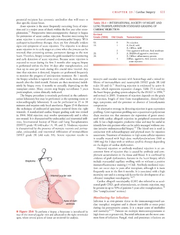
minutes and requires only local anesthesia. Figure 25-8 illustrates compromise.
the technique of endocardial specimen retrieval from the right An alternative strategy in detecting rejection is gene expression
ventricle. A standardized cardiac biopsy grading scale was revised profiling. Gene expression profiling uses a quantitative polymerase
in 2004. Mild rejection may resolve spontaneously and is often chain reaction test that measures the expression of genes associ-
not treated. It is characterized by endocardial and interstitial infil- ated with cardiac allograft rejection in peripheral mononuclear
trate, International Society of Heart and Lung Transplantation cells. It has a high-negative predictive value and may be useful to
(ISHLT) grade 1R (old scales 1A, 1B, and 2). Moderate rejection identify low-risk patients who can be safely managed without rou-
is characterized by the presence of myocyte necrosis and perivas- tine invasive endomyocardial biopsy. 73 The blood test is used in
cular, endocardial, and interstitial infiltration of immunoblasts conjuction with echocardiograpy and physical exam for rejection
ISHLT grade 2R (old scale 3A). Severe rejection results in assessment. Treatment of moderate to high acute cellular rejection
is usually treated with high dose methylprednisolone (500 to
1000 mg) for 3 days with or without cytolitic therapy depending
on the degree of cardiac dysfunction.
Humoral rejection or antibody-mediated rejection is an un-
common form of rejection that is caused by antibody and com-
plement accumulation in the tissue and blood. It is confimed by
evidence of graft dysfunction, features in the heart biopsy, which
Bioptome include myocardial capillary swelling with or without a positive
immunofluorescence staining (
C4d). Antibody-mediated rejec-
tion can occur days to years after transplant, although it is more
frequently seen in the first 6 months. It is associated with a high
mortality rate and is a strong risk factor for the development of ac-
Inter
Int er n n nana jugular vein
nter
al al a a a a a a a a a
al jugu
celerated transplant vasculopathy. 74,75
Cardiac allograft vasculopathy (CAV) also referred to as accel-
erated graft CHD, graft atherosclerosis, or chronic rejection, may
be present in up to 50% of patients 5 years after transplantation. 76
(See “complications” section.)
Monitoring for Infection
Ape
pe
Ape ex x x f f of o o o rig ght Infection is an ever-present threat to the immunosuppressed car-
Ape
x
ex
ve
cle
ve en n n n n ntri ri r tr cle e e diac transplant recipient and is almost inevitable at some point
ve
ve
ve
ve
n
ic
i
c
t
tr
tr
t
during the postoperative course. It is a major cause of morbidity
■ Figure 25-8 To perform a biopsy, a bioptome is introduced by and mortality. 61,77 Patients on multiple immunosuppressants at
way of the internal jugular vein and advanced to the right ventricular high doses are at greater risk. Bacterial infections are the most com-
apex, where several pieces of tissue are retrieved for analysis. mon form of infection. Fungal, viral, and protozoan infections are