
Page 630 - Cardiac Nursing
P. 630
009
009
0/2
6/3
0/2
7:4
5
7:4
1
1
6/3
q
q
q
22.
22.
0
0
0
xd
xd
p
p
A
A
A
ara
ara
t
p
t
06
Pa
g
Pa
5
Pa
e 6
06
e 6
g
g
K34
K34
LWB
LWB
25_
25_
0-c
0-c
LWBK340-c25_ p p pp595-622.qxd 06/30/2009 17:45 Page 606 Aptara
59
5-6
5-6
59
606 PA R T I V / Pathophysiology and Management of Heart Disease
self-injury and prolonged hospitalization. Haloperidol is often Wound infection after CABG surgery occurs despite periopera-
used for sedation. tive antibiotics and aseptic technique. Sternal wound infections
typically present 4 to 14 days after surgery with fever, leukocyto-
Late Postoperative Complications sis, and inflammatory wound with purulent drainage. Sternal
wounds are often associated with a sternal click and sternal insta-
After the fourth postoperative day, most cardiac surgery patients bility. Staphylococci, both Staphylococcus aureus and coagulase-
have short, uncomplicated hospital stays and are discharged to negative staphylococcus, are the most common causative organ-
home. However, postpericardiotomy syndrome, cardiac tampon- ism. 49 Superficial chest wounds are treated with antibiotics and
ade, or incisional wound infection may occur during the last post- local drainage. Deep sternal wounds and mediastinitis are treated
operative period. with surgical débridement and closure or plastic surgical closure
Postpericardiotomy syndrome occurs when traumatized tissue in with muscle flap. The incidence of deep sternal infections range
the pericardial cavity triggers an autoimmune response. Postperi- from 0.25% to 4% and the superficial sternal wound infections
cardiotomy syndrome usually occurs weeks to months after sur- are seen in 2% to 6% after cardiac surgery, both of which prolong
29
gery and results from inflammation of the pleura and pericardium care and increase cost. Infections at the venectomy donor sites
causes aching pericardial pain and severe pleuritic pain. Pleural may also occur and are usually treatable with oral antibiotics, but
and pericardial effusions may accompany the inflammation. severe infections may require open drainage and intravenous an-
Treatment is with ibuprofen, indomethacin, or a brief course of tibiotics.
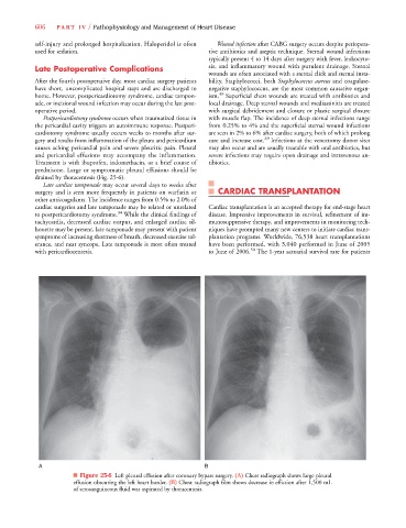
prednisone. Large or symptomatic pleural effusions should be
drained by thoracentesis (Fig. 25-6).
Late cardiac tamponade may occur several days to weeks after
surgery and is seen more frequently in patients on warfarin or CARDIAC TRANSPLANTATION
other anticoagulants. The incidence ranges from 0.5% to 2.0% of
cardiac surgeries and late tamponade may be related or unrelated Cardiac transplantation is an accepted therapy for end-stage heart
34
to postpericardiotomy syndrome. While the clinical findings of disease. Impressive improvements in survival, refinement of im-
tachycardia, decreased cardiac output, and enlarged cardiac sil- munosuppressive therapy, and improvements in monitoring tech-
houette may be present, late tamponade may present with patient niques have prompted many new centers to initiate cardiac trans-
symptoms of increasing shortness of breath, decreased exercise tol- plantation programs. Worldwide, 76,538 heart transplantations
erance, and near syncope. Late tamponade is most often treated have been performed, with 3,040 performed in June of 2005
with pericardiocentesis. to June of 2006. 50 The 1-year actuarial survival rate for patients
A B
■ Figure 25-6 Left pleural effusion after coronary bypass surgery. (A) Chest radiograph shows large pleural
effusion obscuring the left heart border. (B) Chest radiograph film shows decrease in effusion after 1,500 mL
of serosanguineous fluid was aspirated by thoracentesis.