
Page 773 - Cardiac Nursing
P. 773
CHAPTER 31 / Adult Congenital Heart Disease 749
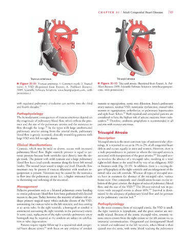
n Figure 31-14 Truncus arteriosus. 1: Common trunk; 2: Truncal n Figure 31-15 Tricuspid atresia. (Reprinted from Everett, A. Ped-
valve; 3: VSD (Reprinted from Everett, A. PedHeart Resource. Heart Resource. 2009. Scientific Software Solutions. www.heartpassport.
2009. Scientific Software Solutions. www.heartpassport.com., with com., with permission.)
permission.)
with regulated pulmonary circulation can survive into the third stenosis or regurgitation, aortic root dilatation, branch pulmonary
and fourth decades. 21 artery stenosis, residual VSD, ventricular dysfunction, truncal valve
stenosis or regurgitation, arrhythmias, or pulmonary hypertension
59
Pathophysiology and right heart failure. Both repaired and unrepaired patients are
The hemodynamic consequences of truncus arteriosus depend on considered to have the highest risk of adverse outcome from endo-
20
the magnitude of pulmonary blood flow, which reflects the pres- carditis. Therefore, antibiotic prophylaxis is recommended in all
ence and the size of the pulmonary arteries and the resistance to patients with truncus arteriosus.
flow through the lungs. 14 In the types with large unobstructed
pulmonary arteries arising from the arterial trunk, pulmonary Tricuspid Atresia
blood flow is greatly increased, clinically resembling patients with
large VSD with left-to-right shunts. Description
Tricuspid atresia is the most common type of univentricular phys-
Clinical Manifestations iology. It is reported to occur in 1% to 2% of all congenital heart
Cyanosis, which may be mild or absent, occurs with increased defects and occurs equally in men and women. However, there is
pulmonary blood flow. Right ventricle pressure is equal to sys- a male preponderance in patients in whom the tricuspid atresia is
62
temic pressure because both ventricles eject directly into the sin- associated with transposition of the great arteries. Tricuspid atre-
gle trunk. The patient with mild cyanosis and a large pulmonary sia involves the absence of a tricuspid valve, resulting in a total
blood flow has a loud systolic murmur along the lower left sternal right-to-left shunt at the atrial level by way of an obligatory ASD
border. The second heart sound is single and loud. Diastolic flow or foramen ovale (Fig. 31-15). In addition, there is a varying de-
murmurs may be present if truncus dilatation with resultant re- gree of hypoplasia of the right ventricle and an enlargement of the
gurgitation is present. Variations may be caused by the resistance mitral valve and left ventricle. Whereas all types of tricuspid atre-
to flow into the pulmonary circuit (i.e., a higher resistance leads sia have in common the absence of the tricuspid valve, various
to shortening and softening of the murmur). forms exist. One commonly used classification focuses on the re-
lation of the great arteries, the degree of reduced pulmonary blood
Management flow, and the size of the VSD. 63 The 20-year survival rate in pa-
Palliative procedures such as a bilateral pulmonary artery banding tients with tricuspid atresia is about 60%. 64 Survival is deter-
to restrict pulmonary blood flow have been performed with limited mined by the adequacy of pulmonary blood flow and the pressure
success in the past. Today, however, the majority of infants will un- in the pulmonary vascular bed. 14
dergo primary surgical repair which includes closure of the VSD,
committing the truncus valve to the left ventricle, and thus serving Pathophysiology
as an aortic valve. In the right ventricle, a homograft or conduit is In the most common form of tricuspid atresia, the VSD is small,
constructed connecting the right ventricle to the pulmonary artery. the right ventricle is hypoplastic, and the great arteries are nor-
In some cases, replacement of the right ventricle–pulmonary artery mally related. Because of the atretic tricuspid valve, systemic ve-
homograft may be required as the conduits are subject to calcifica- nous return crosses from the right atrium to the left atrium via an
tion or valve degeneration. interatrial communication. Oxygenated and deoxygenated blood
Patients require regular follow-up in a specialized adult congen- is mixed and redirected to the left ventricle, where blood is then
ital heart disease center, 26 with focus on any evidence of conduit ejected into the aorta, with some blood reaching the pulmonary