
Page 770 - Cardiac Nursing
P. 770
746 P AR T IV / Pathophysiology and Management of Heart Disease
tricuspid regurgitation. For those who remain asymptomatic, an cyanosis and is referred to as “acyanotic” tetralogy of Fallot. As
annual evaluation that includes ECG and echocardiography is pulmonic obstruction increases, the shunt reverses to right-to-left.
recommended. 26 Endocarditis prophylaxis is not required in the Right ventricular hypertrophy develops due to resistance to the
unrepaired patient, but is recommended in repaired patients with outflow. When total obstruction to pulmonary flow exists, and
prosthetic material or artificial valves. 20 the pulmonary trunk and its branches are present, blood flow to
the lung is mainly through enlarged bronchial arteries and, at
times, also through a PDA. This severe form of tetralogy of Fallot
CYANOTIC HEART DEFECTS WITH is referred to as pulmonary atresia.
DIMINISHED PULMONARY Clinical Manifestations
BLOOD FLOW Typically the patient with tetralogy of Fallot has mild-to-moder-
ate cyanosis and clubbing. The hyperpneic episodes (“hypoxic
Cyanotic heart defects are generally characterized by a right-to-left spells”) associated with tetralogy of Fallot are virtually absent in
shunting, resulting in a flow of desaturated blood into the sys- the adult. The physical appearance is characterized by a small un-
temic circulation. derdeveloped body size. If the patient has only mild cyanosis, de-
velopment is normal. A loud systolic murmur over the third left
Tetralogy of Fallot sternal border with a thrill is characteristic; with severe tetralogy
and marked decrease in pulmonary blood flow the murmur may be
Description short and of low intensity. This is due to the absence of turbulent
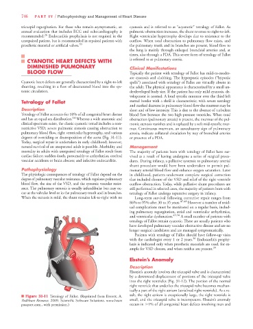
Tetralogy of Fallot accounts for 10% of all congenital heart disease blood flow between the two high-pressure ventricles. When total
14
and has an equal sex distribution. Whereas a wide anatomic and obstruction (pulmonary atresia) is present, the murmur of the pul-
clinical spectrum exists, the classic cyanotic tetrad includes a non- monic stenosis vanishes and is replaced by a soft mid-systolic mur-
restrictive VSD, severe pulmonic stenosis causing obstruction to mur. Continuous murmurs, an auscultatory sign of pulmonary
pulmonary blood flow, right ventricular hypertrophy, and various atresia, indicate collateral circulation by way of bronchial arteries
degrees of overriding or dextroposition of the aorta (Fig. 31-11). or presence of a PDA.
Today, surgical repair is undertaken in early childhood; however,
natural survival of an unoperated adult is possible. Morbidity and Management
mortality in adults with unrepaired tetralogy of Fallot result from The majority of patients born with tetralogy of Fallot have sur-
cardiac failure; sudden death, presumably to arrhythmias; cerebral vived as a result of having undergone a series of surgical proce-
vascular accidents or brain abscess; and infective endocarditis. dures. During infancy, a palliative systemic to pulmonary arterial
shunt procedure would have been undertaken to permit pul-
Pathophysiology monary arterial blood flow and enhance oxygen saturation. Later
The physiologic consequences of tetralogy of Fallot depend on the in childhood, patients underwent complete surgical correction
degree of pulmonary vascular resistance, which regulates pulmonary that included closure of the VSD and relief of the right ventricle
blood flow, the size of the VSD, and the systemic vascular resist- outflow obstruction. Today, while palliative shunt procedures are
ance. The pulmonary stenosis is usually infundibular but may oc- still performed in selected cases, the majority of patients born with
cur at the valvular level or in the pulmonary trunk and its branches. tetralogy of Fallot undergo reparative surgery in infancy.
When the stenosis is mild, the shunt remains left-to-right with no Long-term survival following corrective repair ranges from
86% to 95% after 30 to 35 years. 47,48 However a number of resid-
ual complications must be monitored on a regular basis, includ-
ing pulmonary regurgitation, atrial and ventricular arrhythmias,
and ventricular dysfunction. 49,50 A small number of patients with
tetralogy of Fallot remain cyanotic. These are usually patients who
have developed pulmonary vascular obstructive disease and are no
longer surgical candidates and are managed symptomatically.
Patients with tetralogy of Fallot should have follow-up visits
with the cardiologist every 1 or 2 years. 26 Endocarditis prophy-
laxis is indicated only when prosthetic materials are used, for ex-
ample for VSD closure, and when residua are present. 20
Ebstein’ s Anomaly
Description
Ebstein’s anomaly involves the tricuspid valve and is characterized
by a downward displacement of portions of the tricuspid valve
into the right ventricles (Fig. 31-12). The portion of the normal
right ventricle that underlies the tricuspid valve becomes mechan-
ically a part of the right atrium (atrialized right ventricle). As a re-
n Figure 31-11 Tetralogy of Fallot. (Reprinted from Everett, A. sult, the right atrium is exceptionally large, the right ventricle is
PedHeart Resource. 2009. Scientific Software Solutions. www.heart small, and the tricuspid valve is incompetent. Ebstein’s anomaly
passport.com., with permission.) occurs in .1% of all congenital heart defects involving men and